header image workshops

Single Molecule Workshop

30th International Workshop on “Single Molecule Spectroscopy and Super-resolution Microscopy”

September 23 - 26, 2025, Berlin, Germany

Celebrate 30 years of single molecule research with us!

Image Single Molecule Workshop

News

July 17, 2025
Preliminary program online

We are happy to announce that the preliminary program for the workshop has just been published. For a more detailed overview on the sessions and talks, please refer to our program page

More information >


June 27, 2025
Registration times, end of Workshop and new hotel confirmed

Registration times as well as the end of the workshop have now been confirmed. Registration opens on Tuesday, 23rd September at 8:00 am.

The workshop will end on Friday, 26th September at 5:00 pm.

We are also happy to announce that we will now be cooperating with Nena Apartments for accomodation. For further information on this option, please refer to our accomodation page

More information >


picture of train at Berlin HauptbahnhofMay 6, 2025
Special train fare now available for event attendees

We’re pleased to announce that all participants can now take advantage of a special event ticket offered by Deutsche Bahn. This exclusive offer allows you to travel to our workshop at a discounted rate. Booking is available starting now—don’t miss the chance to save on your journey!

More information >


Interview Tinnefeld on youtubeApril 9, 2025
Video: The full interview with Philip Tinnefeld is now available

From award winner to scientific leader: In an exclusive interview, Prof. Dr. Philip Tinnefeld (LMU Munich), Student Award Winner from 2001, shares his inspiring journey from his early days at the Single Molecule Workshop to becoming a leading expert in single-molecule research. We are happy to have him as an invited speaker at this year's workshop!

Watch the interview on youtube >


April 4, 2025
Media partnership with Wiley's Microscopy and Analysis

We are pleased to announce our media partnership with Microscopy and Analysis. Through this collaboration, we aim to expand the visibility and impact of cutting-edge research in single molecule science.


Former student award winner Philip TinnefeldApril 2, 2025
Where Are They Now? A Look at Past Winners of PicoQuant’s Student Award: An interview with Philip Tinnefeld

Prof. Dr. Philip Tinnefeld (LMU Munich), a 2001 Student Award winner, has maintained a strong connection with the Single Molecule Workshop over the years. This year, he joins once again as an invited speaker. In our exclusive interview, he reflects on his journey, his ongoing ties to the community, and shares insights into the world of single-molecule research.

Read an excerpt from the interview >


Aleksandra Radenovic is invited speakerMarch 27, 2025
Aleksandra Radenovic confirmed as invited speaker

We are happy to confirm Aleksandra Radenovic from the EPFL Lausanne as a further invited speaker at our upcoming anniversary workshop.

Check the list of all invited speakers >


audience single molecule workshopMarch 18, 2025
Last call for abstracts

Don't miss your chance to be part of PicoQuant's 30th Single Molecule Workshop in Berlin!

Abstract deadline: April 16, 2025
PhD students: Submit your abstract for a talk within the framework of our Student Award.

Submit your abstract >


past student award winnersMarch 13, 2025
Where Are They Now? A Look at Past Winners of PicoQuant’s Student Award

As we celebrate 30 years of PicoQuant's Single Molecule Workshop, we’re looking back at the talented young researchers who took the stage and won the Student Award. Read about the inspiring career journeys of past award recipients, from those driving scientific innovation to those leading in industry.

Read more >


poster session single molecule workshopNovember 28, 2024
Registration is open

Registration is now open for our 30th anniversary Workshop on "Single Molecule Spectroscopy and Super-resolution Microscopy". We invite you to submit your abstract for a talk or a poster and join the world's foremost experts in single molecule research.


anniversary iconOctober 17, 2024
Save the date: 30th anniversary workshop September 23–26, 2025

The excitement for next year’s 30th anniversary workshop is already building, promising an extended program with two Nobel laureates and key pioneers in the field. Registration opens in November 2024.

Past workshops

The workshop on "Single Molecule Spectroscopy and Ultra Sensitive Analysis in the Life Sciences" is an annual event since 1995. To get an impression of our Single Molecule Workshops have a look at the video below and check our archive of past events.

Summaries and impressions of past workshops:


Meet the single molecule community in Berlin!

For the 30th year, the world's foremost experts in single molecule research convene in the vibrant city of Berlin. This year's gathering is nothing short of extraordinary. We are honored to host two Nobel laureates, along with the pioneers who paved the way and the rising stars shaping the future.

Audience during an oral presentation

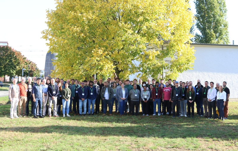
Be part of an exciting and stimulating conference where you can give a talk, present a poster, or attend without presenting. As always, we will award a “Best Student Talk” prize worth 750 Euro.

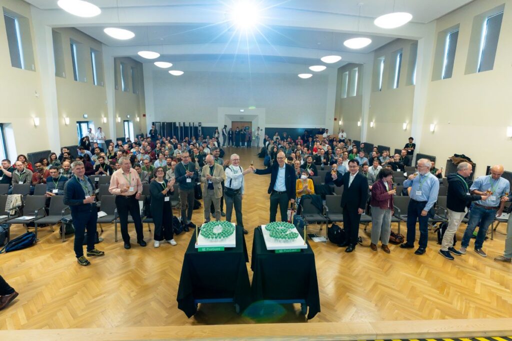
Immerse yourself in a groundbreaking exploration of ultra-sensitive optical detection down to the single molecule level as well as beyond the classical diffraction limit. PicoQuant's renowned workshop provides an unparalleled platform for interdisciplinary collaboration and the exchange of cutting-edge research.

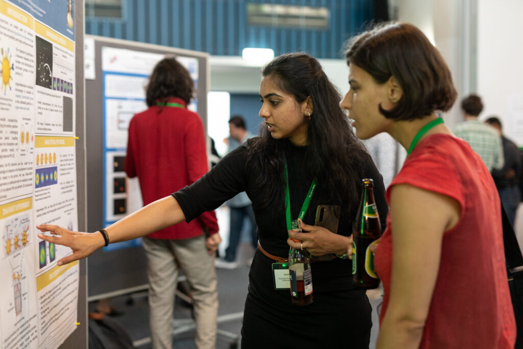
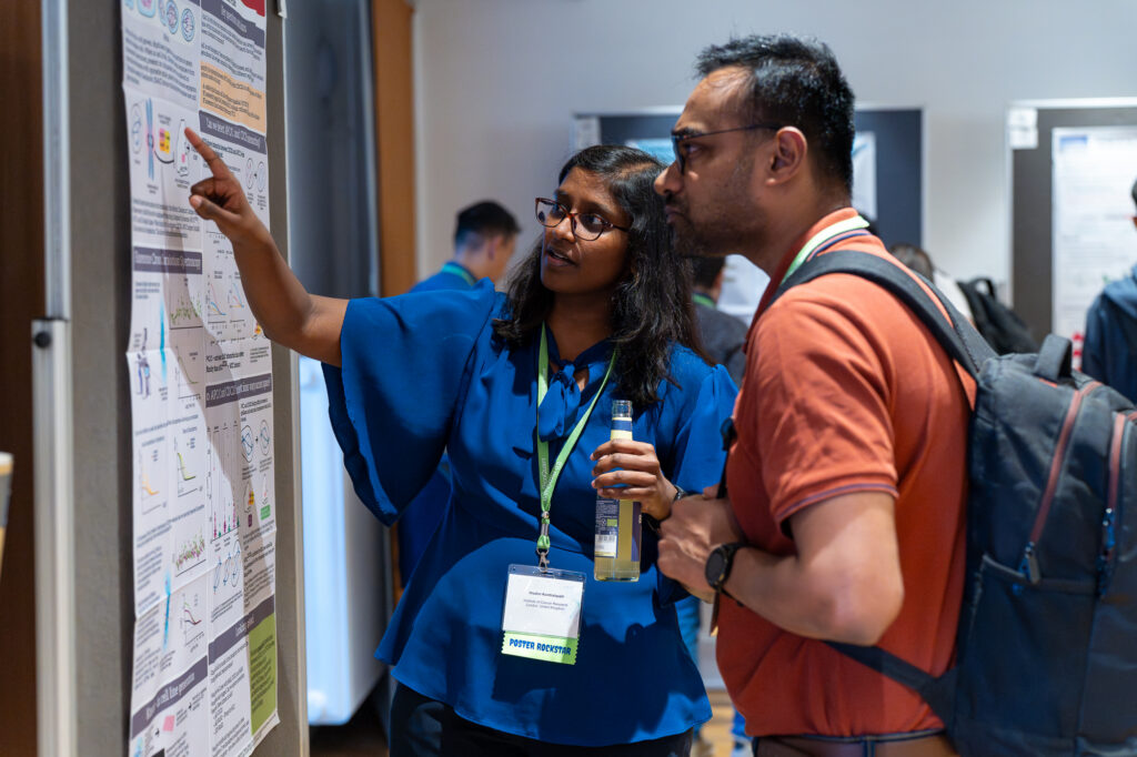
Discover a vast array of applications and methods in single molecule spectroscopy and advanced microscopy, presented through captivating talks and posters. Network with leading experts and like-minded researchers at our welcoming reception and workshop dinner.

Don't miss this opportunity to be at the forefront of scientific innovation!

Covered topics include:

  • Fluorescence Lifetime Imaging (FLIM)
  • Single molecule Förster Resonance Energy Transfer (smFRET)
  • Polarization and Anisotropy based techniques
  • Quantitative imaging methods
  • New fluorescence sensors and labeling schemes
  • Fluorescence Correlation Spectroscopy (FCS) and Fluorescence Lifetime Correlation Spectroscopy (FLCS)
  • Image Scanning Microscopy (ISM)
  • Single Molecule Localization Microscopy (e.g., PALM, STORM, dSTORM, GSDIM, PAINT)
  • Open source data analysis solutions
  • Big Data and Machine Learning approaches to superresolution and single molecule techniques

Both widefield and confocal fluorescence microscopy techniques are covered as well as in vivo and in vitro measurements with single molecule sensitivity.

Important dates

  • Deadline for abstract submission: April 16, 2025
  • Deadline for fee waiver application: April 16, 2025
  • Final deadline for workshop registration August 15, 2025
  • Notification on acceptance of abstracts: August 2025
  • Program available August 2025

Student Award

Icon Student AwardAs nurturing young scientists is important to PicoQuant, we host a competition for the “Best Student Talk” with an award worth 750 Euro. Undergraduate and graduate students are invited to submit their contributions until April 16, 2025. Please indicate during abstract submission if you wish to participate in the contest.


Contact

Workshop coordinators:
Claudia Bergemann
Katharina Zühlke

Tel: +49-30-1208820-87
Email: workshop@picoquant.com

Poster session during the workshop


Microscopy & Analysis logo

Media partner: Microscopy and Analysis
Through our collaboration with Microscopy and Analysis, we aim to expand the visibility and impact of cutting-edge research in single molecule science.

Invited speakers

Guillermo Acuna

Guillermo Acuna
University of Fribourg (UNIFR), Switzerland

Thorben Cordes

Thorben Cordes
TU Dortmund University, Germany

Christian Eggeling

Christian Eggeling
Friedrich Schiller University Jena, Germany

Jörg Enderlein

Jörg Enderlein
University of Göttingen, Germany

Paul French

Paul French
Imperial College London, UK

Maria Garcia-Parajo

Maria Garcia-Parajo
ICFO, Barcelona, Spain

Viktorija Glembockyte

Viktorija Glembockyte
Max Planck Institute for Medical Research, Heidelberg, Germany

Taekjip Ha

Taekjip Ha
Harvard Medical School (HMS), Boston, USA

Mike Heilemann

Mike Heilemann
Goethe University Frankfurt, Germany

Stefan Hell

Stefan Hell (Key Note)
Max Planck Institute for Multidisciplinary Sciences, Göttingen, Germany

Johan Hofkens

Johan Hofkens
KU Leuven, Belgium

Jessica P. Houston

Jessica P. Houston
New Mexico State University,
Las Cruces, USA

Madhavi Krishnan

Madhavi Krishnan
University of Oxford, UK

Don Lamb

Don Lamb
LMU Munich, Germany

W.E. Moerner

W.E. Moerner (Key Note)
Stanford University, USA

Michel Orrit

Michel Orrit
Leiden University, The Netherlands

Aleksandra Radenovic

Aleksandra Radenovic
EPFL, Lausanne, Switzerland

Verena Ruprecht

Verena Ruprecht
Centre for Genomic Regulation (CRG), Barcelona, Spain

Vahid Sandoghdar

Vahid Sandoghdar
Max Planck Society, Erlangen, Germany

Markus Sauer

Markus Sauer
University of Würzburg, Germany

Ben Schuler

Ben Schuler
University of Zurich, Switzerland

Petra Schwille

Petra Schwille
Max-Planck-Institute of Biochemistry, Martinsried, Germany

Claus Seidel

Claus Seidel
Heinrich Heine University Düsseldorf, Germany

Sobhan Sen

Sobhan Sen
Jawaharlal Nehru University, 
New Delhi, India

Allison Squires

Allison Squires
The University of Chicago, USA

Philip Tinnefeld

Philip Tinnefeld
LMU Munich, Germany

Jerker Widengren

Jerker Widengren
KTH Royal Institute of Technology, Stockholm, Sweden

Paul Wiseman

Paul Wiseman
McGill University, Montreal, Canada


Xiaoliang Sunney Xie

Xiaoliang Sunney Xie
Peking University, China


Prelimenary Program

08.00 - 09.00Registration
09.00 - 09.25Rainer Erdmann, Berlin, Germany
Opening Remarks
Session 1: Sensor & LabelChair: Jörg Enderlein
09.25 - 09.50
Thorben Cordes, Dortmund, Germany (Invited Talk)

From accurate FRET studies in proteins to systematic assay design

Thorben Cordes

Biophysical Chemistry, Department of Chemistry and Chemical Biology, Technische Universität Dortmund, Otto-Hahn-Str. 4a, 44227 Dortmund, Germany

Single-molecule FRET (smFRET) has emerged as a powerful tool for studying biomolecular structure and dynamics at the nanoscale. Recent research has focused on improving its reliability and practical applications. Through international blind studies, our community established high measurement precision of quantitative interprobe distances with ≤0.2 nm precision and ≤0.5 nm accuracy [1-2]. While this provided confidence in the use of smFRET for both mechanistic biochemical studies and structural biology, selecting optimal labeling positions for fluorescent dyes remains challenging, particularly in proteins. Empirical guidelines exist for identifying fluorophore labeling sites in proteins, yet, there is no systematic way to predict these site until now. Through literature screening and bioinformatics analysis, we have identified four key parameters that can be combined into a label score system to quantitatively rank residues based on their suitability for fluorophore labelling[3]. We show the predictive power of the score with literature data and new experiments. Available both as a Python script and through a public webserver (https://labelizer.bio.lmu.de/), the Labelizer analyzes protein structures and structural models to predict optimal labeling sites, significantly improving experimental design success rates.


[1] Hellenkamp et al., Nature Methods 15 (2018) 669-676 [2] Agam et al., Nature Methods 20 (2023) 523-535 [3] Gebhardt et al., Nature Communications in press (2025): https://www.biorxiv.org/content/10.1101/2023.06.12.544586.abstract

09.50 - 10.15
Mike Heilemann, Frankfurt am Main, Germany (Invited Talk)

Imaging membrane receptor biology with single-molecule resolution

Mike Heilemann

Institute of Physical and Theoretical Chemistry, Goethe-Universität Frankfurt, Germany

Membrane receptors convert extracellular signals into intracellular responses through highly regulated structural and biochemical mechanisms. Achieving a detailed structural and mechanistic understanding of these signaling processes necessitates observations in living cells. Due to the inherent heterogeneity and lack of synchronization within biological systems, single-molecule experiments are essential. We employ various single-molecule imaging modalities – including quantitative single-molecule localization microscopy (SMLM), single-molecule FRET (smFRET) and single-particle tracking (SPT) – to systematically investigate three key aspects of membrane receptor activation: (i) the molecular stoichiometry and supramolecular assembly patterns of receptor complexes, (ii) the association and dissociation kinetics of receptor complexes, and (iii) the lateral diffusion dynamics of single receptors as a proxy for the microenvironment and activity states. Using these tools and integrating molecular dynamics simulations, we determined the conformation of the receptor-ligand complex (MET:InlB)2 in situ [1] and its dissociation kinetics and lateral mobility in living cells [1, 2]. Furthermore, we established long-term single-particle tracking experiments in living cells and measured ligand-specific activation dynamics of membrane receptor tyrosine kinases [3, 4].


[1] Li, Y., Dietz, M. S., Barth, H. D., Niemann, H. H., Heilemann, M. (2025). Single-molecule FRET-tracking of InlB-activated MET receptors in living cells. bioRxiv, 2025.05. 06.652421. https://www.biorxiv.org/content/10.1101/2025.05.06.652421v1

[2] Li, Y., Arghittu, S. M., Dietz, M. S., Hella, G. J., Haße, D., Ferraris, D. M., Freund, P., Barth, H. D., Iamele, L., de Jonge, H., Niemann, H. H., Covino, R., & Heilemann, M. (2024). Single-molecule imaging and molecular dynamics simulations reveal early activation of the MET receptor in cells. Nature Communications, 15(1), 9486. https://doi.org/10.1038/s41467-024-53772-7

[3] Catapano, C., Rahm, J. V., Omer, M., Teodori, L., Kjems, J., Dietz, M. S., & Heilemann, M. (2023). Biased activation of the receptor tyrosine kinase HER2. Cellular and molecular life sciences: CMLS, 80(6), 158. https://doi.org/10.1007/s00018-023-04806-8

[4] Catapano, C., Dietz, M. S., Kompa, J., Jang, S., Freund, P., Johnsson, K., & Heilemann, M. (2025). Long-Term Single-Molecule Tracking in Living Cells using Weak-Affinity Protein Labeling. Angewandte Chemie International Edition, 64(1), e202413117. https://doi.org/10.1002/anie.202413117

10.15 - 10.30
Patrick Schüler, München, Germany (Student Award)

Beyond strand displacement reactions: DNA computing on the single molecule level

Patrick Schüler, Tim Schröder, Julian Bauer, Philip Tinnefeld

Ludwig-Maximilians-University Munich, Department of Chemistry , Butenandtstr. 5 – 13 (Gerhard-Ertl-Building), D–81377 Munich

DNA computing is an emerging field offering paralleled operation in unconventional media for applications beyond silicon-based computing.[1] Current DNA computing approaches utilize the concept of toehold-mediated strand displacement reactions to create integrated circuits and perform Boolean logic operations. Often these approaches rely on multiple orthogonal diffusive DNA strands, setting limits to sequence space and prolong computation time due to diffusion limited reactions.[2] Recently spatially localized architectures based on the DNA Origami technique could increase calculation time and signal propagation but still require diffusive “fuel” strands for every computational step and was only used in ensemble experiments.[3]

We present a novel DNA computing approach, free of strand displacement reactions. Our confined molecular processing unit (MPU) can perform all essential one- and two-input logic operations. We characterize the MPU performance on the single molecule level as well as read out the calculations of the computation. The single molecule fluorescence readout additionally offers multi-valued logic as readout offering a broad multiplexing potential. Additionally, our MPU is not limited to DNA-inputs but can also be combined with Antibody-Antigen and Protein-Aptamer signal-input for a broad application potential.


[1] M. Adleman, Science 266, 1021-1024 (1994).

[2] G. Seelig, D. Soloveichik, D. Y. Zhang, E. Winfree, Science 314, 1585-1588 (2006).

[3] G. Chatterjee, N. Dalchau, R. A. Muscat, A. Phillips, G. Seelig, Nat Nanotechnol 12, 920-927 (2017).

10.30 - 10.45
Soohyen Jang, Frankfurt am Main, Germany (Student Award)

Quantitative PAINT microscopy of membrane proteins with self-labeling protein tags

Soohyen Jang1,2, Julian kompa3, Claudia Catapano1, Marina S. Dietz1, Kai Johnsson3, Mike Heilemann1,2

1Institute of Physical and Theoretical Chemistry, Johann Wolfgang Goethe-University Frankfurt, Max-von-Laue-Str. 7, 60438 Frankfurt, Germany
2Institute of Physical and Theoretical Chemistry, IMPRS on Cellular Biophysics, Max-von-Laue-Str. 7, 60438 Frankfurt, Germany
3Department of Chemical Biology, Max Planck Institute for Medical Research, Jahnstr. 29, 69120 Heidelberg, Germany

Single-molecule localization microscopy achieves near-molecular resolution by separating the fluorophores in time and space.1 Point accumulation in nanoscale topography (PAINT) separates the fluorescence signal by employing transient binding of fluorophore labels to the target structure1,2. In DNA-PAINT, short DNA oligonucleotides are used to increase the specificity and the number of labeling­ PAINT is free from photobleaching because the fluorophores are continuously supplemented from the imaging buffer.

Next to providing super-resolved images of biomolecules, quantitative PAINT (qPAINT) extracts molecule numbers in tightly packed nano-clusters from the binding kinetics of fluorophores to the target.1,2,4 However, accurate application of qPAINT demands for stoichiometric labeling of target molecules, which is difficult to achieve with widely used antibody labeling. Here, we introduce quantitative PAINT using a self-labeling protein tag, RhoTag1.0, which is targeted transiently by the fluorophore TMR. RhoTag1.0 is used to endogenously label several membrane proteins with a 1:1 ratio to increase the molecular counting precision. We utilized monomeric and dimeric membrane proteins to calibrate quantitative PAINT with the RhoTag1.0. Furthermore, we measured the dimerization of EGFR in resting and EGF-stimulated cells.  


1.    Lelek, M. et al. Single-molecule localization microscopy. Nature Reviews Methods Primers 1, 1–27 (2021).

2.    Sharonov, A. & Hochstrasser, R. M. Wide-field subdiffraction imaging by accumulated binding of diffusing probes. Proc Natl Acad Sci U S A 103, 18911–18916 (2006).

3.    Schnitzbauer, J., Strauss, M. T., Schlichthaerle, T., Schueder, F. & Jungmann, R. Super-resolution microscopy with DNA-PAINT. Nature Protocols 12, 1198–1228 (2017).

4.    Jungmann, R. et al. Quantitative super-resolution imaging with qPAINT. Nat Methods 13, 439–442 (2016).

10.45 - 11.20COFFEE BREAK & EXHIBITION
Session 2: Single Molecule Methods IChair: Petra Schwille
11.20 - 11.45Jörg Enderlein, Göttingen, Germany (Invited Talk)
A journey through 30 years of Single Molecule Science
11.45 - 12.10
Madhavi Krishnan, Oxford, United Kingdom (Invited Talk)

Measurements of molecular size and shape on a chip

Madhavi Krishnan

University of Oxford, United Kingdom

Size and shape are critical discriminators between molecular species and states. I shall describe a microchip-based high-throughput imaging approach offering rapid and precise determination of molecular properties under native solution conditions. Our method detects differences in molecular weight across at least three orders of magnitude and down to two carbon atoms in small molecules. We quantify the strength of molecular interactions across more than six orders of magnitude in affinity constant and track reactions in real time. Highly parallel measurements on individual molecules serve to characterize sample-state heterogeneity at the highest resolution, offering predictive input to model three-dimensional structure. We further leverage the method’s structural sensitivity for diagnostics, exploiting ligand-induced conformational changes in the insulin receptor to sense insulin concentration in serum at the subnanoliter and subzeptomole scale.

12.10 - 12.25
Alexandre Fürstenberg, Geneva, Switzerland

Environment-sensitive fluorescence lifetime probes for single-molecule and super-resolution imaging

Alexandre Fürstenberg

Department of Physical Chemistry and Department of Inorganic and Analytical Chemistry, University of Geneva, Geneva, Switzerland

Despite their importance, fluorescence lifetime imaging (FLIM) and single-molecule-based super-resolution microscopy (SMLM) have rarely been combined into a single experiment, mostly for two reasons: (1) the absence of widely applicable experimental implementation until recently; (2) the sparsity of suitable environment-sensitive probes compatible with both FLIM and SMLM. The experimental issue was solved by the introduction of fluorescence-lifetime single-molecule localization microscopy (FL-SMLM) by the Enderlein group which was followed by a commercially available solution. On the probe side however, although the advent of super-resolution microscopy triggered the development of new fluorescent probes, emphasis was put onto making photostable fluorophores which reliably report their position and whose fluorescence is insensitive to their nanoenvironment.

In this contribution, we describe our efforts to develop targetable probes based on red-emitting fluorophores compatible with SMLM and FLIM whose lifetime directly reports on the number of water molecules in their contact sphere, opening the door to sensing hydration in biological environments such as protein surfaces and microgels [1-2]. In addition, we demonstrate how novel mechanosensitive Flipper probes, which act as membrane tension reporters with broad FLIM applications, can be used in SMLM and single-molecule tracking experiments, enabling to discriminate different membrane compositions at the nanoscale [3-4].


J. Maillard, C. A. Rumble, A. Fürstenberg, Red-Emitting Fluorophores as Local Water-Sensing Probes, J. Phys. Chem. B 125, 9727 (2021).

S. Jana, O. Nevskyi, H. Höche, L. Trottenberg, E. Siemes, J. Enderlein, A. Fürstenberg, and D. Wöll, Angew. Chem. Int. Ed. 63, e202318421 (2024).

J. Garcia-Calvo, J. Maillard, I. Fureraj, K. Strakova, A. Colom, V. Mercier, A. Roux, E. Vauthey, N. Sakai, A. Fürstenberg, S. Matile, Fluorescent Membrane Tension Probes for Super-Resolution Microscopy: Combining Mechanosensitive Cascade Switching with Dynamic-Covalent Ketone Chemistry, J. Am. Chem. Soc. 142, 12034 (2020)

J. Maillard, E. Grassin, E. Bestsennaia, J. Garcia-Calvo, M. Silaghi, N. Sakai, S. Matile, and A. Fürstenberg, J. Phys. Chem. B 128, 7997 (2024)

12.25 - 12.40
Yuval Ebenstein, Tel Aviv, Israel

A Spectral Image Scanning Microscope for Multi-Color Super-Resolution Imaging

Lanna Bram, Yael Roichman, Yuval Ebenstein, Jonathan Jeffe

Raymond and Beverly Sackler Faculty of Exact Sciences, Tel Aviv University, Tel Aviv 6997801, Israel

The recent realization of image scanning microscopy (ISM) using confocal spinning disks (CSD) enhanced the spatial resolution of fluorescence microscopy to twice the diffraction limit with minimal sample perturbation and fast acquisition rates. However, capturing multi-color images using ISM is still time-consuming, and different colors are not acquired simultaneously.  Here, we present a spectral  CSD-ISM system designed for concurrent high-resolution and simultaneous multi-color acquisition. By integrating a custom linear Amici prism into the CSD-ISM optical detection path, we achieve multi-color,  super-resolution images at a fraction of the acquisition time and with a flexible color palette selection.  A digital signal processor (DSP) is employed as a cost-effective alternative to Field-Programmable Gate Arrays (FPGAs) used in previous studies. A GPU-compatible, python-based image processing pipeline decomposes spectral signatures into multi-color images, preserving the optical resolution. System characterization using fluorescent beads demonstrated 1.73-fold resolution improvement over the diffraction limit and accurate color classification with three times faster acquisition compared to standard CSD-ISM. Application to neuron cells induced with Parkinson's disease showcased improved resolution and contrast of four distinctly labeled cellular components. This spectral CSD-ISM system provides a valuable tool for biological imaging, enabling the simultaneous acquisition of high-resolution spatial information and multi-color spectral data.

12.40 - 13.00Flashtalk Session I
13.00 - 14.15LUNCH BREAK
Session 3: Superresolution IChair: Mike Heilemann
14.15 - 14.20Rainer Erdmann, Berlin, Germany
Introduction to Keynote Talk
14.20 - 14.55
Stefan W. Hell, Göttingen, Germany (Invited Talk)

Molecule-scale resolution in dynamics and fluorescence microscopy

Stefan W. Hell & co-workers

Max Planck Institute for Multidisciplinary Sciences, Göttingen & Max Planck Institute for Medical Research, Heidelberg

I will discuss MINFLUX [1-4], a recent molecular localization and superresolution method that has reached Angström localization precision and resolution of the size of a fluorophore molecule. MINFLUX and the related MINSTED concept [5,6] are being established for routine applications in cell and molecular biology, structural biology and neuroscience. Relying on much fewer fluorescence photons than the widely used camera-based localization methods, these techniques are poised to characterize dynamic processes of single proteins, as demonstrated by tracking the nanometer conformational changes of the motor proteins kinesin-1 [7] and dynein in living cells [8]. MINFLUX has also been demonstrated to measure intramolecular distances with Angström precision, providing a precise and reliable alternative to FRET [9]. Harnessing confocal detection, MINFLUX also provides nanometer-range resolution deeper down in layers of cells and (mildly) scattering tissue [10]. Finally, I will show an arguably surprising ability of MINFLUX to separate individual identical fluorophores without sequential ON/OFF switching or activation of fluorescence. Thus, the simultaneous, uninterrupted, nanometer-scale tracking and imaging of multiple, identical (same-color) fluorophores becomes possible for the first time [11]. This novel superresolution principle should allow MINFLUX to reveal the conformational changes of individual proteins in their native environment.


[1] Balzarotti, F., Eilers, Y., Gwosch, K. C., Gynnå, A. H., Westphal, V., Stefani, F. D., Elf, J., Hell, S.W. Nanometer resolution imaging and tracking of fluorescent molecules with minimal photon fluxes. Science 355, 606-612 (2017).

[2] Eilers, Y., Ta, H., Gwosch, K. C., Balzarotti, F., Hell, S. W. MINFLUX monitors rapid molecular jumps with superior spatiotemporal resolution. PNAS 115, 6117-6122 (2018).

[3] Gwosch, K. C., Pape, J. K., Balzarotti, F., Hoess, P., Ellenberg, J., Ries, J., Hell, S. W. MINFLUX nanoscopy delivers 3D multicolor nanometer resolution in cells. Nat. Methods 17, 217–224 (2020).

[4] Schmidt, R., Weihs, T., Wurm, C. A., Jansen, I., Rehman, J., Sahl, S. J., Hell, S. W. (2021) MINFLUX nanometer-scale 3D imaging and microsecond-range tracking on a common fluorescence microscope. Nat. Commun. 12:1478.

[5] Weber, M., Leutenegger, M., Stoldt, S., Jakobs, S., Mihaila, T. S., Butkevich, A. N., Hell, S. W. MINSTED fluorescence localization and nanoscopy. Nat. Photon. 15, 361-366 (2021).

[6] Weber, M., von der Emde, H., Leutenegger, M., Gunkel, P., Sambandan, S., Khan, T. A., Keller-Findeisen, J., Cordes, V. C., Hell, S.W. MINSTED nanoscopy enters the Ångström localization range. Nat. Biotechnol., 41, 569-576 (2023).

[7] Wolff, J. O., Scheiderer, L., Engelhardt, T., Engelhardt, J., Matthias, J., Hell, S.W. MINFLUX dissects the unimpeded walking of kinesin-1. Science, 379, 1004-1010 (2023).

[8] Schleske, J. M., Hubrich, J., Wirth, J. O., D’Este, E., Engelhardt, J., Hell, S. W. MINFLUX reveals dynein stepping in live neurons. PNAS 121, e2412241121 (2024).

[9] Sahl, S. J., Matthias, J., Inamdar, K., Weber, M., Khan, T. A., Brüser, C., Jakobs, S., Becker, S., Griesinger, C., Broichhagen, J., Hell, S. W. Direct optical measurement of intramolecular distances with angstrom precision. Science 386, 180-187 (2024).

[10] Moosmayer, T., Kiszka, K. A., Pape, J. K., Leutenegger, M., Steffens, H., Grant, S. G. N., Sahl, S. J., Hell, S. W. MINFLUX fluorescence nanoscopy in biological tissue. PNAS 121, e2422020121 (2024).

[11] Hensel, T. A., Wirth, J. O., Schwarz, O. L. Hell, S. W. Diffraction minima resolve point scatterers at few hundredths of the wavelength. Nat. Phys. https://doi.org/10.1038/s41567-024-02760-1 (2025).

14.55 - 15.20
Aleksandra Radenovic, Lausanne, Switzerland (Invited Talk)

Advancing Single-Molecule Imaging

Aleksandra Radenovic

EPFL, Lausanne, Switzerland

In this talk, I will demonstrate how advancements in imaging methods can open new avenues for research at solid-liquid interface. In the first part, I will discuss our efforts to explore nanophotonics at the single-molecule level, with a focus on the optical dynamics and conformations of individual emitters at the solid-liquid interface and in confined environments. In our recent work, we employed fluorescence microscopy to monitor electrochemical reactions at the single-molecule scale, achieving a wide field of view and high temporal resolution. Additionally, we introduced an advanced bifocal polarization single-molecule localization microscopy (pSMLM) to enable real-time, multi-dimensional observations of quantum emissions formed by organic molecules and h-BN native defects. Our findings reveal a strong correlation between the orientation of quantum emitters and the symmetry of the h-BN lattice. I will also discuss the use of SPAD cameras for single-particle tracking applications, as well as in high-throughput single-molecule fluorescence lifetime imaging (smFLIM).


[1] Ronceray, Nathan, Yi You, Evgenii Glushkov, Martina Lihter, Benjamin Rehl, Tzu-Heng Chen, Gwang-Hyeon Nam et al. "Liquid-activated quantum emission from pristine hexagonal boron nitride for nanofluidic sensing." Nature Materials 22, no. 10 (2023): 1236-1242.

[2] Mayner, Eveline, Nathan Ronceray, Martina Lihter, Tzu-Heng Chen, Kenji Watanabe, Takashi Taniguchi, and Aleksandra Radenovic. "Monitoring electrochemical dynamics through single-molecule imaging of hBN surface emitters in organic solvents." arXiv preprint arXiv:2405.10686 (2024).

[3] Guo, Wei, Tzu-Heng Chen, Nathan Ronceray, Eveline Mayner, Kenji Watanabe, Takashi Taniguchi, and Aleksandra Radenovic. "Dipole orientation reveals single-molecule interactions and dynamics on 2D crystals." arXiv preprint arXiv:2408.01207 (2024).

15.20 - 15.35
Rick Seifert, Würzburg, Germany (Student Award)

Correlative confocal and super-resolution imaging of the immunological CAR-T cell synapse

Rick Seifert1, Leon Gehrke2, Nicole Seifert1, Sören Doose1, Michael Hudecek2, Thomas Nerreter2, Markus Sauer1

1Department of Biotechnology and Biophysics, Biocenter, University of Würzburg, Würzburg, Germany
2Chair in Cellular Immunotherapy, Medical Clinic II, University Hospital of Würzburg, Würzburg, Germany

Chimeric antigen receptor (CAR)-T cell therapy uses T cells engineered to express CARs specifically targeting surface antigens on cancer cells. Upon recognition, CAR-T cells form immunological synapses (IS) against cancer cells and eliminate them. Unlike the conventional bull’s-eye IS of T cells, CAR-T cells form a multifocal IS. The ultrastructure of this IS can be used for assessing the efficacy of different CAR designs [1].

To investigate and quantify CAR rearrangement during IS formation, we are developing a correlative confocal and super-resolution microscopy approach. For 3D quantification of CARs, we developed a deformable mirror-based 3D-direct stochastic optical reconstruction microscopy (dSTORM) workflow. The deformable mirror allows us to engineer a tetrapod point spread function (PSF). Additionally, we integrated a Re-scan confocal microscope (RCM) [2], enabling correlative RCM and 3D-dSTORM imaging. This allows us to correlate CAR distribution at a high spatial resolution with different cellular structures and other membrane receptors.

Our approach provides new insights into CAR-T cell IS architecture and offers a potential framework to evaluate CAR designs based on IS ultrastructure for improved CAR-T cell immunotherapies.


[1] Liu D, Badeti S, Dotti G, et al., Cell Communication and Signaling, 18(1), 134 (2020)

[2] De Luca GMR, Breedijk RMP, Brandt RAJ, et al., Biomed Opt Express, 4(11), 2644 (2013)

15.35 - 15.50
Francisco Matos, Orsay, France (Student Award)

TimeLoc: Integrating Dynamic Excitation and SPAD units for Camera-Free Frequency-Encoded Super-Resolution Imaging and Tracking

Francisco Matos1, Maximilian Lengauer1,2, Emmanuel Fort2, Sandrine Lévêque-Fort1

1Institut des Sciences Moléculaires d Orsay, 598 Rue André Rivière, 91400 Orsay, France
2Institut Langevin,1 Rue Jussieu, 75005 Paris, France

Fluorescence super-resolution microscopy capabilities have progressively advanced by leveraging molecular sparsity to achieve nanometer-scale localization. While conventional methods typically rely on camera-based detection and spatial PSF fitting, recent approaches have turned toward temporal encoding to enhance localization performance [1].

Pursuing this line of development, Time Localization Microscopy (TimeLoc) employs dynamic structured excitation, encoding each position in the field of view with a distinct temporal modulation frequency [2]. This frequency-based strategy enables spatial localization through analysis of the emitted signal, removing the need for pixelated camera detection by using a Single Photon Avalanche Diode (SPAD).

Initial experiments with a single SPAD have demonstrated the feasibility of wide-field localization based solely on temporal modulation, highlighting the potential of frequency-based encoding for spatial reconstruction. Building on these results, a 23-element SPAD array is now being incorporated to enable parallel detection and expand spatial sampling capacity.

Current efforts focus on evaluating array-based performance and assessing two-dimensional excitation schemes, including sequential and simultaneous modulation modes. These studies aim to improve the robustness of frequency encoding and support scalable imaging and tracking. Preliminary results using dSTORM and DNA-PAINT will be presented as part of the continued assessment of the technique.


[1] Jouchet, P; Cabriel, C.; Bourg, N.; et al.: Nanometric axial localization of single fluorescent molecules with modulated excitation. Nature Photonics, Vol. 15, 2021, pp. 297–304. DOI: 10.1038/s41566-020-00749-9.

[2] Lengauer, M: Wide-field super-resolution imaging from a time-modulated fluorescence signal. Dissertation, Université Paris-Saclay and École Supérieure de Physique et de Chimie Industrielles de la Ville de Paris, 2023.

15.50 - 16.10Flashtalk Session II
16.10 - 16.40COFFEE BREAK & EXHIBITION
Session 4: Biological ApplicationsChair: Aleksandra Radenovic
16.40 - 17.05
Petra Schwille, Martinsried, Germany (Invited Talk)

Understanding biology by building it? The exciting world of synthetic cells.

Petra Schwille

Max-Planck-Institute of Biochemistry, Martinsried, Germany

tba

17.05 - 17.30
Verena Ruprecht, Innsbruck, Austria (Invited Talk)

Mechano-signalling as a regulator of cell behaviour 

Verena Ruprecht

University of Innsbruck, Austria

tba

17.30 - 17.45
Abhilash Kulkarni, Stockholm, Sweden (Student Award)

Time-gated detection of NIR luminescent nanoparticles in organs using snSPDs

Abhilash Kulkarni1, Olof Eskilson2,3, Georgios A. Sotiriou2,3, Jerker Widengren1

1Experimental Biomolecular Physics, Dept. of Applied Physics, Albanova University Center, Royal Institute of Technology (KTH), 106 91 Stockholm, Sweden.
2Department of Microbiology, Tumor and Cell Biology, Karolinska Institutet, 171 17, Stockholm, Sweden.
3Department of Materials and Environmental Chemistry, Stockholm University, 106 91, Stockholm, Sweden.

Luminescent nanoparticles (NPs) are promising candidates for near-infrared (NIR) in-vivo and biomedical imaging due to their high brightness, photostability, and low photobleaching. Their surface can be functionalized with proteins to target specific tissues. This study investigates the biodistribution of Neodymium (Nd)-doped NPs in vital mouse organs, exploring their potential as drug carriers for both diagnostic and therapeutic applications.

We utilize Superconducting Nanowire Single Photon Detectors (snSPDs), previously shown to offer high time resolution, low dark counts and no afterpulsing in NIR single-molecule studies(1). The current snSPD is optimized for detecting sharp NP emission lines in the 900–1100 nm range, improving penetration depth and reducing autofluorescence and scattering in biological media.

We exploit techniques pioneered in single molecule spectroscopy to enhance the quantitative information gained to accurately estimate the biodistribution. We take advantage of long excited state lifetime of the NPs to perform time gated detection with a high repetition rate laser combined with confocal laser scanning microscopy at various depths. Burst analysis is performed to differentiate actual emission from noise. Furthermore, we implement a coincidence count-based detection to improve SBR by employing a Hanbury-Brown-Twiss arrangement. Results are benchmarked against ICP-MS and compared with an in-house camera-based lock-in detection method.(2)


(1) Kulkarni, A.; Bagheri, N.; Widengren, J. Multiplexed Near-IR Detection of Single-Molecule Fluorescence Fluctuations Using a Single Superconducting Nanowire Single-Photon Detector. ACS Photonics 2025, 12 (4), 2233-2241. DOI: 10.1021/acsphotonics.5c00224.

(2) Bagheri, N.; Wang, C.; Guo, D.; Lakshmanan, A.; Zhu, Q.; Ghazyani, N.; Zhan, Q.; Sotiriou, G. A.; Liu, H.; Widengren, J. Lanthanide upconversion nonlinearity: a key probe feature for background-free deep-tissue imaging. 2025; p arXiv:2503.05325.

17.45 - 18.00
Tom Kache, Diepenbeek, Belgium (Student Award)

Unraveling DNA Ligase Dynamics via Multiparameter Photon-by-Photon smFRET and H2MM Analysis

Tom Kache1, David M Wilson III2, Jelle Hendrix1

1Dynamic Bioimaging Lab, BIOMED, Hasselt University, 3590 Diepenbeek, Belgium
2The GRAND Team, BIOMED, Hasselt University, 3590 Diepenbeek, Belgium

Multiparameter, photon-by-photon, single-molecule FRET (smFRET) allows for integrating several aspects of fluorescence detection to aid the structural study of biomolecules. Here, we employed anisotropy-resolved smFRET and mpH2MM to characterize the interplay between FRET-labeled nicked DNA (nDNA) and purified human DNA ligase 3 (LIG3). LIG3 stands out among human DNA ligases as the sole mitochondrial DNA ligase and the only one possessing an additional DNA-binding zinc-finger domain capable of sensing DNA nicks. smFRET allowed us to detect discrete LIG3-induced bending states of the nDNA substrate and an increase in anisotropy, indicative of tight protein-DNA interactions. By maintaining constant ionic strength, we observed that varying magnesium concentrations modulated the nDNA FRET and anisotropy signatures, suggesting different binding modes for LIG3. Employing anisotropy-resolved multiparameter H2MM analysis, we identified several structural states that dynamically interconvert at rates of hundreds of times per second. The rate and nature of these conformational changes were found to depend on the presence of magnesium ions. Our results align with previous structural and kinetic studies that have suggested a dynamic hand-off mechanism between the zinc-finger and the catalytic part of the protein, which may regulate LIG3's function.

18.00 - 18.15
Marie Reischke, Erlangen, Germany (Student Award)

Chip-based iSCAT microscopy under evanescent illumination

18.15 - 18.20VOTING STUDENT AWARD
18.20 - 19.50POSTER SESSION I & GET TOGETHER
Session 5: Single Molecule Methods IIChair: Paul French
09.00 - 09.25
Xiaoliang Sunney Xie, Beijing, China (Invited Talk)

tba

Xiaoliang Sunney Xie

Peking University, China

tba

09.25 - 09.50
Paul Wiseman, Montréal, Canada (Invited Talk)

Pollen Tube Growth Dynamics Quantified via Volumetric Spatio-Temporal Image Correlation Spectroscopy

Paul Wiseman

McGill University, Montreal, Canada

Rapid pollen tube growth is key for fertilization from pollen to the ovary in flowering plants. Tube growth necessitates the expansion of adjoining cell walls and plasma membranes, both requiring local delivery of the new material via cargo vesicles which contain precursor molecules within a lipid bilayer. Due to the three-dimensional planar form of a cell wall, cellular expansion must be precisely controlled in space with targeted deposition of new material to specific surface areas of the growing cell front. Biophysical characterization of pollen tube growth in space and time is challenging due to the high density of transport vesicles and requires a combination of high-temporal-frequency 3D imaging and advanced image analysis methods. 3D imaging time series of growing pollen tubes were collected via field-synthesis lattice light-sheet microscopy to minimize photobleaching and increase sample viability for long-term imaging. To quantify vesicle trafficking, the volumetric extension of the established 2D spatio-temporal image correlation spectroscopy (STICS) was used to provide the first 3D mapping of vectorial transport of the cargo vesicles in a living plant pollen tube. We also performed lifetime-filtering FLIM, to separate fluorescence signal localized to vesicles from mitochondrial-localized emission to simultaneously measure cargo vesicle and mitochondrial transport dynamics via STICS, and the local redox microenvironment in growing pollen tubes from camellia japonica pollen grains

09.50 - 10.05
Marcelle Koenig, Berlin, Germany

Expanding the Horizon of FCS with SPAD Arrays: A Promising Outlook for New Applications

Marcelle Koenig1, Anders Barth1, Evangelos Sisamakis1, Fabian Barachati1, Johan Hummert1, Felix Koberling1, Ivan Michel Antolovic2, Rainer Erdmann1

1PicoQuant GmbH, Rudower Chaussee 29, 12489 Berlin, Germany
2Pi Imaging Technology SA, EPFL Innovation Park, 1015 Lausanne, Switzerland

Fluorescence Correlation Spectroscopy (FCS) is a well-established tool for studying molecular interactions and dynamics at the single-molecule level. The recent integration of single-photon avalanche diode (SPAD) arrays combined with time-resolved instrumentation in confocal microscopy provides new possibilities for FCS that provide new insights for live cell investigations.

Here, we evaluate enhanced FCS applications which are enabled by the integration of a cooled high-performance 23-pixel SPAD-array that was developed jointly with Pi Imaging Technologies as an add-on to the confocal microscope Luminosa. The SPAD array allows for the simultaneous detection of multiple fluorescence signals based on single photon counting with high temporal resolution. Any pixel combination within the SPAD array can selected for advanced FCS analyses. Thus, compared to point detectors, spatially resolved information about molecular diffusion and dynamics becomes available. This enables e.g. spot-variation FCS for the identification of potentially hindered diffusion in live cell investigations. Spatial pixel cross-correlations can be used to uncover directional diffusion. The integration of Time-Correlated Single-Photon Counting (TCSPC) provides further information about the fluorescence lifetimes. These can be utilized for an even more comprehensive understanding of complex biological mechanisms.

The integration of SPAD-arrays with time-resolved detection represents a significant advancement for confocal microscopes. Apart from the improved optical resolution for imaging purposes via image scanning microscopy (ISM), SPAD array based detection allows for a multitude of new FCS modalities for studying complex biological processes in both temporal and spatial domains.

10.05 - 10.20
Dominic A. Helmerich, Würzburg, Germany

Unveiling the invisible: A novel approach illuminates the sub-10 nm cosmos

Dominic A. Helmerich1, Made Budiarta2, Danush Taban1, Mara Meub1, Marcel Streit2, Alexander Kuhlemann1, Sören Doose1, Gerti Beliu2, Markus Sauer1,2

1Department of Biotechnology and Biophysics, Biocenter, University of Würzburg, Am Hubland, 97074, Würzburg, Germany
2Rudolf Virchow Center, Research Center for Integrative and Translational Bioimaging, University of Würzburg, 97080, Würzburg, Germany

Super-resolution microscopy has transformed our understanding of cellular structures, achieving spatial resolution down to single-digit nanometers. Despite advancements in angstrom-level accuracy on reference structures, applying this precision to biological samples remains difficult. Our work presents a novel approach that turns this challenge into an opportunity for discovery.

Here we utilize photoswitching fingerprint analysis, which leverages the unique temporal behavior of fluorescent dyes to extract information in the sub-10 nm range. By analyzing the blinking patterns of fluorophore systems, we can reveal molecular arrangements and interactions that were previously hidden.

This method combines advanced analysis with specially designed protein-based reference structures, enabling precise calibration of super-resolution techniques. The stability of these structures in cellular environments makes them ideal for benchmarking high-resolution imaging methods and avoiding misinterpretations during live cell studies.

This innovative methodology opens new avenues for exploring the nanoscale world of biology, providing unprecedented insights into cellular processes and molecular interactions.


D. A. Helmerich, G. Beliu, D. Taban, et al., Nat Methods, 19, 986–994 (2022)

D. A. Helmerich, M. Budiarta, et al., Adv. Mater., 36, 2310104 (2024)

10.20 - 10.35
Tao Chen, Göttingen, Germany

Measuring membrane and membrane protein structure and dynamics with dynamic metal- and graphene-induced energy transfer spectroscopy (dynaMIET/dynaGIET)

Narain Karedla, Jörg Enderlein, Tao Chen

Georg-August-Universitat Gottingen, Drittes Physikalisches Institut-Biophysik, Friedrich-Hund-Platz 1, 37077 Göttingen, GERMANY

Cell membranes are dynamic, fluid structures composed of a phospholipid bilayer with embedded proteins, exhibiting fluidity and viscoelastic properties essential for various biological functions like cell signaling, membrane trafficking, and cell division.  However, accurately measuring the dynamics of intricate membrane systems, like mitochondria, characterized by rapid and subtle fluctuations, poses significant challenges. In this study, we introduce a novel methodology (dynaMIET/dynaGIET) that combines metal/graphene-induced energy transfer (MIET/GIET) (1) with various fluorescence correlation spectroscopy (FCS)- based techniques to precisely quantify the structure and dynamics of different membrane systems. With these combinations, we measured the membrane fluctuations (2), leaflet-specific structure and diffusions (3, 4), and membrane protein conformation dynamics (5). Moreover, we showcase the versatility and applicability of dynaMIET/dynaGIET in studying various membrane systems. 


1. A. I. Chizhik, J. Rother, I. Gregor, A. Janshoff, J. Enderlein, Metal-induced energy transfer for live cell nanoscopy. Nat. Photonics 8, 124–127 (2014).

2. T. Chen, N. Karedla, J. Enderlein, Measuring sub-nanometer undulations at microsecond temporal resolution with metal- and graphene-induced energy transfer spectroscopy. Nat. Commun. 15, 1789 (2024).

3. N. Karedla, F. Schneider, J. Enderlein, T. Chen, Leaflet-specific Structure and Dynamics of Solid and Polymer Supported Lipid Bilayers. Angew. Chem. Int. Ed. n/a, e202423784.

4. T. Chen, A. Ghosh, J. Enderlein, Cholesterol-Induced Nanoscale Variations in the Thickness of Phospholipid Membranes. Nano Lett. 23, 2421–2426 (2023).

5. T. Chen, N. Karedla, J. Enderlein, Observation of E-cadherin adherens junction dynamics with metal-induced energy transfer imaging and spectroscopy. Commun. Biol. 7, 1–10 (2024).

10.35 - 10.45GROUP PICTURE
10.45 - 11.20COFFEE BREAK & EXHIBITION
Session 6: Superresolution IIChair: Michel Orrit
11.20 - 11.25Rainer Erdmann, Berlin, Germany
Introduction to Keynote Talk
11.25 - 12.00
W.E. Moerner, Stanford, United States (Invited Talk)

A Brief Survey of Single-Molecule Optical Microscopy: From Early Spectroscopy in Solids, to Super-Resolution Nanoscopy in Cells, to a Wealth of New Applications

W. E. (William E.) Moerner

Departments of Chemistry and Applied Physics (courtesy) Stanford University, Stanford, CA USA 94305

First observed optically 36 years ago in my laboratory at IBM Research, single molecules have enabled much interesting science spanning physics, chemistry, materials science, medicine, and biology. In this new field of optical microscopy of the nanoscale, ensemble averaging is removed, so each single molecule can act as a reporter of not only its position, but also about local information about the nearby environment. Combined with blinking and photoswitching (first observed at low temperatures in 1992 and then for single GFP proteins at room temperature in 1997) to ensure sparsity, in the mid-2000’s, super-resolution fluorescence microscopy based on single molecules has opened up a frontier in which structures and behavior can be observed in materials and in fixed and live cells with resolutions down to the one and two digit nm scale. Cellular studies of single molecules have been enhanced by PSF engineering to extract 3D position and orientation, deep learning to estimate molecular variables and structured backgrounds, light sheet illumination, and much more. A recent study shows fascinating intracellular structures formed by SARS-CoV-2 viral RNA and proteins in infected mammalian cells. Three-dimensional single-molecule tracking in live cells provides time-dependent information about biological regulation and condensed complexes, as well as about anomalous diffusion of DNA loci in nuclei and more. Acronyms abound (PAINT, ODMR, ABEL,…) - the future is bright, indeed!

12.00 - 12.25
Jörg Enderlein, Göttingen, Germany (Invited Talk)

Advancing Super-Resolution Imaging: Integrating Fluorescence Lifetime, Scanning Microscopy, and Energy Transfer Techniques for Isotropic Nanoscale Bioimaging

Jörg Enderlein

Georg August University Göttingen, Germany

Recent advancements in super-resolution microscopy have enabled unprecedented insights into the spatial organization of cellular structures. In this talk, I will present a series of methodological innovations that synergistically integrate fluorescence-lifetime single-molecule localization microscopy (FL-SMLM) [1,2], image scanning microscopy (ISM) [3,4], and metal- or graphene-induced energy transfer (MIET/GIET) imaging [5–7]. These approaches collectively offer isotropic three-dimensional resolution at the nanometer scale, multiplexed imaging capabilities, and robustness against chromatic aberrations. First, I will discuss our work on MIET and GIET microscopy, which exploit distance-dependent quenching phenomena near metallic or graphene interfaces to determine the axial position of single emitters with sub-10 nm accuracy. The combination of MIET with direct Stochastic Optical Reconstruction Microscopy (dSTORM) or DNA-based Points Accumulation for Imaging in Nanoscale Topography (DNA-PAINT) provides truly isotropic 3D resolution, extending the reach of localization microscopy into the axial dimension without interferometric complexity. Second, I will highlight the development of fluorescence lifetime DNA-PAINT (FL-PAINT), a technique that enables multi-target super-resolution imaging through fluorescence lifetime multiplexing without fluid exchange. By utilizing orthogonally designed imager strands conjugated to fluorophores with distinct lifetimes, we achieve simultaneous imaging of multiple targets in the dense intracellular environment. Lastly, I will introduce our latest development of fluorescence-lifetime image scanning microscopy SMLM (FL-iSMLM), which achieves a near twofold enhancement in lateral resolution by integrating a single-photon detector array into a confocal laser scanning microscope (CLSM). This method combines the localization precision of ISM with the multiplexing power of fluorescence-lifetime detection, enabling sub-5 nm resolution in fixed cells while simultaneously allowing discrimination of targets based solely on their fluorescence lifetimes.


[1] J. C. Thiele, D. A. Helmerich, N. Oleksiievets, R. Tsukanov, E. Butkevich, M. Sauer, O. Nevskyi, and J. Enderlein, Confocal Fluorescence-Lifetime Single-Molecule Localization Microscopy, ACS Nano 14, 14190 (2020).

[2] J. C. Thiele, O. Nevskyi, D. A. Helmerich, M. Sauer, and J. Enderlein, Advanced Data Analysis for Fluorescence-Lifetime Single-Molecule Localization Microscopy, Front. Bioinform. 1, 740281 (2021).

[3] C. B. Müller and J. Enderlein, Image Scanning Microscopy, Phys. Rev. Lett. 104, 198101 (2010).

[4] N. Radmacher, O. Nevskyi, J. I. Gallea, J. C. Thiele, I. Gregor, S. O. Rizzoli, and J. Enderlein, Doubling the resolution of fluorescence-lifetime single-molecule localization microscopy with image scanning microscopy, Nat. Photon. 18, 1059 (2024).

[5] A. I. Chizhik, J. Rother, I. Gregor, A. Janshoff, and J. Enderlein, Metal-induced energy transfer for live cell nanoscopy, Nature Photon 8, 124 (2014).

[6] A. Ghosh, A. Sharma, A. I. Chizhik, S. Isbaner, D. Ruhlandt, R. Tsukanov, I. Gregor, N. Karedla, and J. Enderlein, Graphene-based metal-induced energy transfer for sub-nanometre optical localization, Nat. Photonics 13, 860 (2019).

[7] J. C. Thiele, M. Jungblut, D. A. Helmerich, R. Tsukanov, A. Chizhik, A. I. Chizhik, M. Schnermann, M. Sauer, O. Nevskyi, and J. Enderlein, Isotropic Three-Dimensional Dual-Color Super-Resolution Microscopy with Metal-Induced Energy Transfer, Science Advances 8, 14190 (2021).

12.25 - 12.50
Guillermo Acuna, Fribourg, Switzerland (Invited Talk)

Direct single-molecule detection and super-resolution imaging with a low-cost portable smartphone-based microscope

Guillermo Acuna

Department of Physics, University of Fribourg, Switzerland

We present a novel, low-cost, portable smartphone-based fluorescence microscope capable of detecting single-molecule fluorescence directly, i.e., without the need for any signal amplification. The setup leverages the image sensors and data handling capacity of mass-produced smartphones, making it adaptable to different smartphones and capable of detecting single molecules across the visible spectral range. We showcase this capability through single-molecule measurements on DNA origami models and super-resolution microscopy of biological cells by single-molecule localization microscopy. Last, we illustrate its potential as a point-of-care (POC) device by implementing a single-molecule bioassay for RNA detection. This development paves the way for biotechnology innovations, making use of massively distributed or personalized assays with single-molecule sensitivity, with the potential to revolutionize digital bioassays, POC diagnostics, field expeditions, STEM outreach, and life science education.

12.50 - 13.05
Christian Franke, Jena, Germany

Nanotexture – a universal approach of AI-based computational multiplexing and phenotyping of super-resolution data.

Bela Vogler1,2, Gregor Gentsch1, Pablo Caravilla1,2,3, Dominic Helmerich4, Teresa Klein4, Katharina Reglinski1,2, Markus Sauer4,5, Christian Eggeling1,2, Christian Franke1

1Faculty of Physics and Astronomy, Institute of Applied Optics and Biophysics, Friedrich Schiller University Jena, Jena, Germany
2Leibniz Institute of Photonic Technology e.V., Jena, Germany, member of the Leibniz Centre for Photonics in Infection Research (LPI), Jena, Germany
3Present address: Science for Life Laboratory, Department of Women’s and Children’s Health, Karolinska Institutet, Solna, Sweden
4Department of Biotechnology and Biophysics, Biocenter, University of Würzburg, Am Hubland, Würzburg, Germany
5Rudolf Virchow Center, Research Center for Integrative and Translational Bioimaging, University of Würzburg, Würzburg, Germany

Fluorescence-based super-resolution microscopy (SRM) enables nanometer-scale visualization of cellular organelles. Traditional multi-color SRM relies on spectral multiplexing, but leading methods—STED, SMLM, MINFLUX—require specialized dyes with delicate photo-physical properties, limiting multi-color imaging fidelity and live-cell compatibility. Fast acquisition techniques like SIM and Airy-Scan also face speed-resolution trade-offs and lack synchronicity in multi-color applications.

We introduce NanTex, a ML-based context-agnostic multiplexing approach leveraging organelle-specific nanotextures, applicable to SMLM, MINFLUX, STED, SIM, and Airy scan microscopy. NanTex demixes overlapping organelles from single-channel images without spectral separation, using AI-enabled textural demixing via U-Net learning [1].

NanTex trained on SMLM is directly applicable to MINFLUX without retraining, facilitating multiplexing at MINFLUX resolution without the extreme task to gather sensible amounts of training data. We demonstrate multiplexing in artificial overlays and real experimental datasets with almost all major cellular organelles (actin, microtubules, clathrin, endosomes, lysosomes, ER, mitochondria, golgi, etc.), including live-cell SIM and airyscan multiplexing. Furthermore, we present NanTex on our novel high-speed SIM, based on random pattern structured illumination (speckle SIM) with 100 nm resolution at framerates of >100 Hz [2].

NanTex also enables computational phenotyping, exemplified by quantifying microtubule depolymerization upon nocodazole treatment.

NanTex advances super-resolution multi-organellar imaging in diverse SRM techniques.


[1] B. Vogler, G.J. Gentsch et al. in preparation (will be on biorxiv in February 2025)

[2] A. Platz, G.J. Gentsch et al. in preparation (will be on biorxiv in April 2025)

13.05 - 13.15
Steffen J. Sahl, Göttingen, Germany

Ångström-level, intra-molecular MINFLUX analyses of protein conformation and chemical architecture

13.15 - 14.35LUNCH BREAK
Session 7: Instrumentation & SoftwareChair: Jörg Enderlein
14.35 - 15.00
Michel Orrit, Leiden, Netherlands (Invited Talk)

Looking back at 35 years of single-molecule optics 

Michel Orrit

Leiden University, Netherlands

Beyond scanning probe microscopies, a variety of optical signals give direct access to single molecules and single nanoparticles. For more than 30 years, fluorescence has been the workhorse of single-molecule optics, and has spawned breakthroughs in optical superresolution. A large variety of photochemical and biochemical processes influence fluorescence and thereby give access to the ultimate single-molecule level, free from ensemble averaging. More recently, under the lead of several groups, other optical techniques have reached single-molecule sensitivity. Photothermal microscopy proved sensitive enough to detect single photostable dye molecules or single organic conjugated polymers. The differential absorption of circularly polarized light provides quantitative circular dichroism data of single absorbing chiral or magnetic nanoparticles. Non-absorbing nanoparticles and large molecules can now be detected individually through their optical polarizability only, without need for fluorescent or absorbing labels. Their selectivity and signal-to-noise ratio are enhanced considerably in the near-field of plasmonic gold nanoparticles or even in wide field, thanks to resonant optical microcavities. The arrival and departure of single protein molecules from a solution cause sudden steps in the optical signal, opening micro-analytical applications and in-situ sensing. The capacity to detect and characterize single unlabeled diffusing protein molecules on-the-fly opens fascinating perspectives in bio-medical science.

15.00 - 15.25
Vahid Sandoghdar, Erlangen, Germany (Invited Talk)

Coherent scattering of light by single molecules

Vahid Sandoghdar

1Max Planck Institute for the Science of Light, 91058 Erlangen, Germany
2Max-Planck-Zentrum für Physik und Medizin, 91054 Erlangen, Germany 3Department of Physics, Friedrich-Alexander University of Erlangen-Nürnberg, 91058 Erlangen, Germany

The very first report of single-molecule detection in 1989 involved cryogenic extinction measurements assisted by double-FM modulation [1]. This achievement was followed by experiments, which were conducted based on the detection of the Stokes-shifted fluorescence. However, lack of coherence in spontaneously emitted fluorescence hampered access to the phase of the molecular wavefunction and of the laser light. In 2007, we revisited direct extinction measurements on single molecules with a simple realization that the intrinsic extinction cross section is large enough to be detected directly if the illumination laser field is confined strongly [2]. A series of near-field [3] and far-field [4,5] experiments demonstrated extinction dips as large as 20%. Such a large efficiency in the interaction between light and a single quantum system opened the door to nonlinear effects at the few-photon level [6]. Furthermore, combination with high-finesse microcavities opened the door to near-unity extinction, strong coupling and single-photon nonlinearity [7,8]. More recently, these developments were used to show that coherent cooperative coupling of several individual molecules via a common mode of a microcavity [9]. Interestingly, coherent Rayleigh scattering has also become a powerful method for label-free detection and tracking of biological nanoparticles and nanostructures such as cellular organelles and viruses, down to single proteins at ambient conditions [10]. In this lecture, I will give an overview of these developments and discuss some exciting new perspectives.


[1] W. E. Moerner and L. Kador, PRL 62, 2535 (1989).

[2] G. Zumofen, N. Mojarad, V. Sandoghdar, M. Agio, PRL 101, 180404 (2008).

[3] I. Gerhardt, G. Wrigge, P. Bushev, G. Zumofen, R. Pfab, V. Sandoghdar, PRL 98, 033601 (2007)

[4] G. Wrigge, I. Gerhardt, J. Hwang, G. Zumofen, V. Sandoghdar, Nat. Phys. 4, 60 (2008).

[5] M. Potoschnig, Y. Chassagneux, J. Hwang, G. Zumofen, A. Renn, V. Sandoghdar, PRL 107, 063001 (2011).

[6] A. Maser, B. Gmeiner, T. Utikal, S. Götzinger, V. Sandoghdar, Nat. Photonics 10, 450 (2016).

[7] D. Wang, H. Kelkar, D.-M. Cano, D. Rattenbacher, A. Shkarin, T. Utikal, S. Götzinger, V. Sandoghdar, Nat. Physics 15, 483 (2019).

[8] A. Pscherer, M. Meierhofer, D. Wang, H. Kelkar, D.-M. Cano, T. Utikal, S. Götzinger, V. Sandoghdar, PRL 127, 133603 (2021).

[9] J. Nobakht, A. Pscherer, J. Renger, S. Götzinger, V. Sandoghdar, PNAS, June 2025.

[10] N. Ginsberg, C-L. Hsieh, P. Kukura, M. Piliarik, V. Sandoghdar, Nat. Rev. Meth. Prim. 5, 23 (2025).

15.25 - 15.50
Paul French, London, United Kingdom (Invited Talk)

openScopes: an open, modular platform to widen access and capabilities in microscopy and high content analysis

Paul French

Imperial College London, United Kingdom

I will review recent development and applications of our multidimensional fluorescence and phase imaging instrumentation and outline how we are working to widen access to such advanced imaging modalities, including for high content analysis (HCA) through our “openScopes” open instrumentation platform (www.openscopes.com). openScopes includes research-grade instruments for multidimensional fluorescence and quantitative phase imaging, super-resolved microscopy, and optical projection tomography. To widen access, we have developed an open-source modular, microscope stand, “openFrame” that can be used flexibly for affordable and sustainable instruments in lower resource (e.g., LMIC) settings or for rapidly prototyping of advanced microscopy concepts. A basic openFrame-based brightfield/fluorescence microscope (from ~£10,000) can be upgraded to almost any other microscopy modality. For HCA and slide scanning (e.g., for histopathology), we have developed a novel open-source optical autofocus module. Advanced openFrame-based instruments include automated SMLM based on easySTORM, and a modular multiphoton multiwell plate microscope (M3M) incorporating multiplexed TCSPC FLIM and quantitative phase imaging using our single-shot polarisation differential phase contrast (pDPC) technique that enables label-free single cell segmentation and tracking for long time-lapse fluorescence (FRET) assays.

15.50 - 16.05
Kunihiko Ishii, Wako, Japan

Independent component analysis disentangles fluorescence signals from diffusing single molecules

Kunihiko Ishii1,2, Miyuki Sakaguchi1, Tahei Tahara1,2

1Molecular Spectroscopy Laboratory, RIKEN, 2-1 Hirosawa, Wako 351-0198, Japan
2Ultrafast Spectroscopy Research Team, RIKEN Center for Advanced Photonics (RAP), RIKEN, 2-1 Hirosawa, Wako 351-0198, Japan

Single-molecule fluorescence (SMF) measurement, including smFRET, has become an essential tool in current biophysical research, serving as a unique probe for molecular interactions and structural heterogeneities. By applying SMF to freely diffusing molecules, one can resolve and characterize subpopulations in solutions, making it a promising approach for studying molecules in complex environments. Multiparameter fluorescence detection (MFD) is gradually being adopted in SMF. MFD incorporates additional information into SMF from measurements using fluorescence lifetime, multicolor excitation/detection, and fluorescence anisotropy. Though MFD mitigates the limitation of SMF due to signal weakness by offering enhanced information content, a general framework for data analysis has not yet been established to fully utilize its potential. We recently discovered that independent component analysis (ICA) is ideally suited for analyzing MFD-based SMF data. In this work, we developed an algorithm of ICA optimized for MFD data analysis named IFCA [1]. Applications to static and dynamic mixture systems demonstrate its potential allowing model-free separation of subpopulations with microsecond time resolution in nanomolar concentration regime. To promote its usage, we implemented the developed algorithm with Python and made it compatible with common photon data formats [2].


[1] K. Ishii, M. Sakaguchi, T. Tahara, bioRxiv, https://doi.org/10.1101/2025.03.04.641393 (2025).

[2] K. Ishii, M. Sakaguchi, T. Tahara, Zenodo, https://doi.org/10.5281/zenodo.14869092 (2025).

16.00 - 16.15
Andriy Chmyrov, Heidelberg, Germany

Imaging Beyond The Visible: advantages of the Shortwave-Infrared spectral range for confocal microscopy and Raman scattering imaging.

16.15 - 16.30
Markus Lippitz, Bayreuth, Germany

Fluorescence-detected two-dimensional electronic spectroscopy of a single molecule

Sanchayeeta Jana, Simon Kehrer, Markus Lippitz

Experimental Physics III, University of Bayreuth, Germany

Single-molecule fluorescence spectroscopy is a powerful method that avoids ensemble averaging, but its temporal resolution is limited by the fluorescence lifetime to nanoseconds at most. At the ensemble level, two-dimensional spectroscopy provides insight into ultrafast femtosecond processes such as energy transfer and line broadening, even beyond the Fourier limit, by correlating pump and probe spectra. Here, we combine these two techniques and demonstrate coherent 2D spectroscopy of individual dibenzoterrylene (DBT) molecules at room temperature [1]. We excite the molecule in a confocal microscope with a phase-modulated train of femtosecond pulses and detect the emitted fluorescence with single-photon counting detectors. Using a phase-sensitive detection scheme, we were able to measure the nonlinear 2D spectra of most of the DBT molecules we studied. Our method is applicable to a wide range of single emitters and opens new avenues for understanding energy transfer in single quantum objects on ultrafast time scales.


[1[ S. Jana, S. Durst, M. Lippitz, Nano Letters, 24, 12576-12581 (2024)

16.30 - 23.00SOCIAL PROGRAM & DINNER
Session 8: Biological Applications IChair: Claus Seidel
09.00 - 09.25
Christian Eggeling, Jena, Germany (Invited Talk)

A route through studying molecular membrane dynamics - an advanced microscopy story

Christian Eggeling

Friedrich-Schiller-Universität Jena, Germany

tba

09.25 - 09.50
Jerker Widengren, Stockholm, Sweden (Invited Talk)

Fluorophore blinking in superresolution microscopy, and as a rich source of moleular-scale information

Jerker Widengren

Department of Applied Physics, Royal Inst of Technology (KTH), Stockholm, Sweden

Reversible dark state transitions of fluorophores are central for all forms of fluorescence-based, single-molecule and super-resolution microscopy and spectroscopy, both as limiting factors and as prerequisites. An additional aspect of such transitions is that they also can be used to sense a manifold of biomolecular environments, dynamics and interactions. To make monitoring of dark state transitions widely applicable for studies on biological samples, we have developed so-called transient state (TRAST) imaging [1]. In TRAST, fluorophore dark state transitions are monitored via the time-averaged fluorescence intensity, and from how it varies with the modulation of the excitation light. Here, recent work within the H2020 project NanoVIB will be presented where TRAST is used to characterize dark state transitions in near-IR cyanine fluorophores, how such transitions can compromise MINFLUX super-resolution imaging, and how this knowledge allowed us to formulate strategies to overcome these problems and make near-IR MINFLUX imaging possible [2]. Also, examples will be given, demonstrating how biologically relevant environmental and molecular interaction parameters can be monitored via fluorophore dark state transitions, in solutions, live cells, and tissues, which are difficult, if possible at all, to follow via regular fluorescence readout parameters. Acknowledgements: European Commission (H2020, NanoVIB, 101017180), Swedish Research Council (VR), Swedish Foundation for Strategic Research (SSF).


[1] Widengren, J. in Fluorescence Spectroscopy and Microscopy in Biology, Springer 2023 (Sachl, R., Amaro, M.M. Eds)

[2] Venugopal Srambickal, C. Esmaeeli H. et al., in revision 2025, BioRXiv: 10.1101/2024.08.27.609859

09.50 - 10.05
Gaukhar Zhurgenbayeva, Jena, Germany (Student Award)

Characterization of Candidalysin peptide self-aggregation on lipid membranes using Fluorescence Correlation Spectroscopy.

10.05 - 10.20
Stijn Dilissen, Diepenbeek, Belgium (Student Award)

Dynamic Burst smFRET in Slow Motion: A Microfluidic Approach for Probing Biocondensates and Liposomes

Stijn Dilissen1,2, Pedro L. Silva1, Tom Kache1, Ronald Thoelen2,3, Jelle Hendrix1

1UHasselt, Dynamic Bioimaging Lab, Advanced Optical Microscopy Centre and Biomedical Research Institute, Agoralaan C (BIOMED), B3590 Diepenbeek, Belgium
2UHasselt, Biomedical Device Engineering group, Institute for Materials Research (IMO-IMOMEC), Wetenschapspark 1, B3590 Diepenbeek, Belgium
3IMOMEC Division, IMEC vzw, Wetenschapspark 1, B3590 Diepenbeek, Belgium

Burst-wise single-molecule FRET (smFRET) is a well-established technique for studying protein dynamics in solution, where molecules diffuse through the confocal volume. However, applying smFRET on slow-diffusing molecules within macromolecular assemblies, such as liposomes or biomolecular condensates formed by liquid-liquid phase separation (LLPS), is challenging. These embedded molecules diffuse slowly, and LLPS condensates often sink in solution, making Brownian diffusion-based smFRET less effective. As a result, only a small fraction of the sample is detected, and the longer burst durations increase the risk of photobleaching.

To address these challenges, we introduce a microfluidic chip that delivers precisely controlled, slow flows just above the diffusion rate of these macromolecular assemblies. By maintaining laminar flow, a more significant fraction of the sample can be efficiently probed. The adjustable flow rate allows fine-tuning burst durations: faster flows enable rapid sampling at higher laser powers. In comparison, slower flows extend bursts, enhancing temporal resolution and capturing slower molecular dynamics. Beyond flow control, the chip allows for on-chip reagent mixing or the introduction of conformational change triggers.

Altogether, this microfluidic platform enables a new way to investigate single-molecule behaviour within macromolecular assemblies like liposomes or LLPS condensates, offering deeper insight into cellular organisation and function.

10.20 - 10.35
Robert B. Quast, Montpellier, France

Dissecting the GPCR conformational landscape using biorthogonal click chemistries and multicolor single molecule FRET

Léo Bonhomme1, Ecenaz Bilgen2, Nathalie Lecat-Guillet3, Hongkang Liu3, Caroline Clerté1, Jean-Philippe Pin3, Philippe Rondard3, Don C. Lamb2, Emmanuel Margeat1, Robert B. Quast1

1Centre de Biologie Structurale (CBS), Univ. Montpellier, CNRS, INSERM, Montpellier, France.
2Department of Chemistry, Ludwig-Maximilians-Universität München (LMU), Munich, Germany.
3Institut de Génomique Fonctionnelle, Univ. Montpellier, CNRS, INSERM, 141 rue de la Cardonille, 34094, Montpellier Cedex 05, France.

G protein-coupled receptor (GPCR) activation is mediated by a complex interplay of conformational changes. To decipher the conformational landscape of the human metabotropic glutamate receptors 2 (mGluR2), we developed several conformational 2-color FRET sensors through incorporation of a single[1] and two distinct, reactive ncAAs combined with bioorthogonal conjugation of donor and acceptor fluorophores. We then studied ligand induced conformational rearrangements of single receptors in a carefully optimized detergent mixture[2] by smFRET using confocal, pulsed interleaved excitation and multiparameter fluorescence detection. We find that the natural full agonist glutamate is sufficient to close the Venus flytrap domains. In contrast a synergy with positive allosteric modulators, acting over a long-range functional link, is required to fully activate these multidomain neuroreceptors. Finally, using 3-color smFRET sensors, by combining double ncAA and SNAP-tag labeling, we provide evidence for a previously unknown, pre-active, intermediate state in equilibrium with the active state upon ligand activation that we could not resolve by classical 2-color smFRET[3]. We conclude an activation model where orthosteric and allosteric ligands act on different conformational equilibria between coexisting states to fine-tune mGluR2 activation. Our study highlights the power of minimally invasive, ncAA-based, bioorthogonal labeling to dissect domain-specific conformational rearrangements of GPCRs using smFRET.


[1] Quast RB*, Lecat-Guillet N*, Liu H, Bourrier E, Møller TC, Rovira X, Soldevila S, Lamarque L, Trinquet E, Liu J, Pin JP, Rondard P, Margeat E. Sci. Adv. 9, eadf1378 (2023), https://doi.org/10.1126/sciadv.adf1378

[2] Quast RB*, Cao AM*, Fatemi F, Rondard P, Pin JP, Margeat E. Nat Commun 12, 5426 (2021), https://doi.org/10.1038/s41467-021-25620-5

[3] Bonhomme L, Bilgen E, Clerté C, Pin JP, Rondard P, Margeat E, Lamb DC, Quast RB. bioRxiv 2024, https://doi.org/10.1101/2024.10.31.621373

10.35 - 11.10COFFEE BREAK & EXHIBITION
Session 9: FLIM, FRET & FCS IChair: Viktorija Glembockyte
11.10 - 11.35
Jessica P. Houston, Las Cruces, United States (Invited Talk)

Applications of fluorescence lifetime measurements in flow cytometry

Jessica P. Houston

Jessica P. Houston, Ph.D., Chemical & Materials Engineering, New Mexico State University, Las Cruces, NM 88003, USA

Methods for high throughput single-cell analyses have become quite complex over the last decade with emerging technologies that advance the speed of imaging and sorting as well as enhance the number of parameters that can be measured from a single cell. Many instruments, cytometers, or similar devices provide essential features about cells because optical measurements provide not only spatial but also temporal information about the intracellular environ-ment. Time-resolved flow cytometry (TRFC) is one form of cytometry that captures temporal information about fluo-rescent molecules inside the cell. Such information does not rely on brightness and often correlates to signaling events, molecular movement, and dynamics of molecular interaction. Various TRFC technologies will be presented as well as applications that focus on metabolic mapping of tamoxifen resistant breast cancer cells using autofluorescence. Focus will also be placed on a chip-based cytometer that utilizes acoustic focusing for more accurate fluorescence life-time measurements. The long-term impact of this work is to develop new tools that provide more quantitative fluo-rescence information at the throughput of a flow cytometer.

11.35 - 12.00
Claus Seidel, Düsseldorf, Germany (Invited Talk)

FRET nanoscopy maps molecules of life

Claus Seidel

Heinrich Heine University Düsseldorf, Germany

Multimodal fluorescence spectroscopy and microscopy with multiparameter detection provide rich insights on biomolecular systems with respect to structural and kinetic properties and spatial localization. Single-molecule FRET experiments offer the required nanosecond time resolution to study the motions biomolecules over a large dynamic time range from nano- to milliseconds [1,2]. If more than two FRET states are present, an appropriate analysis of a kinetic network is challenging. Thus, I introduce parametric relations (FRET-lines) [3,4] between two FRET indicators, the donor fluorescence lifetime and the intensity-based FRET efficiency, that significantly facilitate an interpretation. Model-based FRET-lines serve as pathfinders to decode the dynamic spectroscopic fingerprint by: (i) identifying the number of conformational states, (ii) resolving their dynamic connectivity, (iii) comparing different kinetic models, and (iv) inferring polymer properties of unfolded or intrinsically disordered proteins. We assessed the accuracy of this approach in a comparative single-molecule study [5] using two protein systems with distinct conformational changes and dynamics. We obtained an interdye distance precision of ≤2 Å and accuracy of ≤5 Å. Finally, I introduce a framework for FRET nanoscopy with seamless resolution accessing distances from µm down to 5 nm with a precision < 0.7 nm. To enable dissemination of all results according to the FAIR principle, we introduce the flrCIF data representation, which extends established data standards from the Protein Data Bank and allows for archiving fluorescence-aided integrative structures for multiple states together with associated kinetic data on exchange in the PDB-Dev repository.


[1] Sisamakis, E. et al.; Methods Enzymol. 475, 455-514 (2010). doi.org/10.1016/S0076-6879(10)75018-7.

[2] Lerner, E. et al; eLife 10, e60416 (2021). doi.org/10.7554/eLife.60416

[3] Barth, A. et al.; J. Chem. Phys. 156, e141501 (2022). doi.org/10.1063/5.0089134

[4] Opanasyuk, O. et al.; J. Chem. Phys. 156, e031501 (2022). doi.org/10.1063/5.0095754

[5] Agam, G. et al.; Nat. Methods 20, 523–535 (2023). doi.org/10.1038/s41592-023-01807-0

12.00 - 12.15
Léa Brito, Orsay, France (Student Award)

Instant FLIM in SMLM via SPAD Array Imaging

Léa Brito, Laurent Le, Lancelot Pincet, Sandrine Lévêque-Fort

Institut des Sciences Moléculaires d'Orsay, 598 Rue André Rivière, 91400 Orsay, France

Single Molecule Localization Microscopy provides a unique access to 3D nanometric cellular organization. Beyond this gain in spatial information, complementary parameters such as fluorescence lifetime imaging (FLIM) also represents a major interest for a detection at the single molecule level. This approach enables not only the identification of various dyes, but also the probing of the cellular environment. This can be achieved through time gating, where photons are collected only during a defined time window, enabling precise lifetime measurements but which should also preserve the photon budget and associated precision. As sequential gating can lead to a loss of fluorophores which turn ON-OFF stochastically, alternative faster implementation is needed.  

A  new technology, a 512 x 512 SPAD array  initially designed for classical wide field FLIM, enables time-gated imaging with a high signal-to-noise ratio compatible with single molecule detection. Our detection module, developed in combination with this large SPAD array, enables the simultaneous acquisition of two temporal gates. This implementation allows for the concurrent reconstruction of SMLM and ratio-FLIM images over a 50 × 25 µm field of view. A complete characterization of the Instant FLIM system, along with demonstrations on biological samples, will be presented.

12.15 - 12.30
Philipp Gebauer, Konstanz, Germany (Student Award)

Investigating Spectral Fluctuations in the Emission of Halide-Perovskite Nanoparticles using Heralded Spectroscopy

Philipp Gebauer1, Frieder Conradt1, Chenglian Chu2, Eva Haage1, Ihor Cherniukh2, Claudio Bruschini3, Gabriele Rainò2, Edoardo Charbon3, Maksym Kovalenko2, Alfred Leitenstorfer1, Ron Tenne4

1Department of Physics and Center for Applied Photonics, University of Konstanz, D-78464 Konstanz, Germany
2Institute of Inorganic Chemistry, Department of Chemistry and Applied Biosciences, ETH Zürich, CH-8093 Zürich, Switzerland
3School of Engineering, École Polytechnique Fédérale de Lausanne (EPFL), Neuchâtel 2002, Switzerland
4Schulich Faculty of Chemistry, Technion-Israel Institute of Technology, 3200003 Haifa, Israel

Thanks to their bright and tunable emission, colloidal nanoparticles are strong candidates for use in modern electro-optical applications and biological imaging. Halide-perovskite nanoparticles have recently emerged as a promising candidate for generating quantum states of light [1]. However, instabilities in the spectrum of emitted light remain a major challenge, significantly hindering their practical integration into future quantum systems [2].
To better understand the mechanisms behind these spectral instabilities, we employ Heralded Spectroscopy - a recently developed method to measure spectrally resolved photon correlations [3]. It enables the detection of single-photon events with high temporal (~ 100 ps) and spectral (~ 0.2 meV) resolution using a Single-Photon Avalanche Diode (SPAD) array. We use this setup to measure spectral fluctuations of individual nanoparticles under cryogenic conditions. This enables us to extend the statistical analysis to visually trace spectral fluctuations across more than ten orders of magnitude in the time domain, down to nanoseconds. We find a critical dependence of the magnitude of spectral fluctuations on the laser excitation power. Surprisingly, we observe a power-induced transient increase in the lattice symmetry. To understand this exciting finding, we intend to perform a systematic investigation of particles with varying sizes and shapes.


[1] Kaplan, A.E.K., Krajewska, C.J., Proppe, A.H. et al., Nat. Photon. 17, 775-780 (2023).

[2] F. Conradt, V. Bezold, V. Wiechert, S. Huber, S. Mecking, A. Leitenstorfer and R. Tenne, Nano
Lett., 23, 21 (2023)

[3] G. Lubin, R. Tenne, A. C. Ulku, I. M. Antolovic, S. Burri, S. Karg, V. J. Yallapragada, C. Bruschini,
E. Charbon and D. Oron, Nano. Lett., 21, 16 (2021)

12.30 - 12.45
Chi-Jui Feng, Bethesda, United States

Characterizing Barrier Crossing Dynamics of Protein Folding Through Transition Paths Using Single-molecule FRET in Zero-mode Waveguides

Chi-Jui Feng, Ulrich Baxa, John M Louis, Hoi Sung Chung

Laboratory of Chemical Physics, National Institute of Diabetes and Digestive and Kidney Diseases, National Institutes of Health, Bethesda, Maryland 20892, United States

Transition paths (TPs) describe short-lived, single-molecule barrier crossing events that encode conformational dynamics of the molecular transitions, including folding and binding of proteins and nucleic acids. Single-molecule spectroscopy has been hailed to study properties associated with the TPs including TP times around 10–100 μs, free energy landscape along one-dimensional distance coordinate, and diversity of the TPs. However, in single-molecule FRET (smFRET), limited brightness impedes characterization of the TPs around microsecond or shorter. To enhance the brightness to study protein folding TPs, we combined smFRET spectroscopy with zero-mode waveguides. Waveguides successfully enhanced the brightness to several MHz to study microsecond TPs. We surveyed several proteins with varied sizes, secondary structures, and folding rates to discuss relations between various physicochemical properties, kinetics, and barrier crossing dynamics. We found a strong determinant of the TP times is curvature modulated by the barrier height, rather than number of native contacts or size of proteins. We also found that diffusivity of native contact formation empirically scales with the absolute contact order, which is a manifestation of kinetic cooperativity in contact formation. We expect this approach serves as an effective modality to study fast kinetics and dynamics in general folding and binding processes.

12.45 - 13.05Flashtalk Session III
13.05 - 14.20LUNCH BREAK
Session 10: FLIM, FRET & FCS IIChair: Jessica Houston
14.20 - 14.45
Viktorija Glembockyte, Heidelberg, Germany (Invited Talk)

Leveraging DNA Nanotechnology for Single-Molecule Optical Sensing

Viktorija Glembockyte

Max Planck Institute for Medical Research, Heidelberg, Germany

Optical labels and sensors are invaluable tools for understanding biological processes and dissecting disease mechanisms. The advent of single-molecule and super-resolution imaging techniques has placed increasingly stringent demands on these probes, necessitating high photostability for labels and single-molecule sensitivity with high optical contrast for sensors. This presentation will detail our recent efforts to address these challenges by combining single-molecule imaging with DNA nanotechnology. Specifically, we utilize DNA origami to decouple sensing from signal output, enabling the creation of modular and tunable sensor platforms that exhibit both large Förster Resonance Energy Transfer (FRET) contrast and single-molecule sensitivity (Nat. Nanotech. 2025, 20, 303). The inherent modularity of this DNA origami approach allows for the development of single-molecule sensors targeting diverse analytes, including nucleic acids, antibodies, and enzymes, simply by exchanging sensing elements. Furthermore, the incorporation of multiple sensor elements facilitates cooperativity, tunable dynamic range, and advanced logic sensing operations. While DNA nanotechnology holds immense promise for biomedical applications, its widespread utility is currently limited by the inherent instability of DNA nanostructures within complex biochemical environments. We will also discuss our ongoing research aimed at understanding and enhancing the addressability and functionality of DNA nanodevices, leveraging the power of single-molecule and super-resolution imaging (Adv. Mater. 2023, 35, 2212024; Small 2025, in press).

14.45 - 15.10
Ben Schuler, Zurich, Switzerland (Invited Talk)

Probing rapid biomolecular dynamics with single-molecule spectroscopy

Ben Schuler

Universität Zürich, Switzerland

Single-molecule spectroscopy enables biomolecular dynamics to be investigated across more than twelve orders of magnitude in time1, which allows us to probe the molecular mechanisms of a wide range of biological processes. By combining single-molecule FRET with nanosecond FCS, dynamics in the submicrosecond range can be resolved, even in complex environments, such as biomolecular condensates2 and live cells3. I will illustrate recent advances in probing the nanosecond dynamics of disordered proteins4, 5 and nucleic acids6, the increasing synergy with molecular simulations, and the accuracy with which distance distributions and dynamics in such systems can be obtained7.


1. Nettels D., Galvanetto N., Ivanovic M. T., Nüesch M., Yang T. J., Schuler B. Single-molecule FRET for probing nanoscale biomolecular dynamics. Nat. Rev. Phys. 6, 587-605 (2024).

2. Galvanetto N., Ivanovic M. T., Chowdhury A., Sottini A., Nuesch M. F., Nettels D., . . . Schuler B. Extreme dynamics in a biomolecular condensate. Nature 619, 876-883 (2023).

3. König I., Soranno A., Nettels D., Schuler B. Impact of In-Cell and In-Vitro Crowding on the Conformations and Dynamics of an Intrinsically Disordered Protein. Angew. Chem. Int. Ed. 60, 10724-10729 (2021).

4. Nüesch M. F., Ivanovic M. T., Claude J. B., Nettels D., Best R. B., Wenger J., Schuler B. Single-molecule Detection of Ultrafast Biomolecular Dynamics with Nanophotonics. J. Am. Chem. Soc. 144, 52-56 (2022).

5. Chowdhury A., Nettels D., Schuler B. Interaction Dynamics of Intrinsically Disordered Proteins from Single-Molecule Spectroscopy. Annu. Rev. Biophys. 52, 433-462 (2023).

6. Nüesch M. F., Pietrek L., Holmstrom E. D., Nettels D., von Roten V., Kronenberg-Tenga R., . . . Schuler B. Nanosecond chain dynamics of single-stranded nucleic acids. Nat. Commun. 15, 6010 (2024).

7. Nüesch M., Ivanovic M. T., Nettels D., Best R. B., Schuler B. Accuracy of distance distributions and dynamics from single-molecule FRET. Biophys. J., in press (2025).

15.10 - 15.25
Jakob Hartmann, München, Germany (Student Award)

Investigation of DNA and DNA-Protein-Interaction on the Nanometer Scale using Graphene Energy Transfer

Jakob Hartmann1, Lars Richter1, Giovanni Ferrari1, Alan Szalai1, Merve-Zeynep Kesici1, Bosong Ji1, Izabela Kamińska1,2 and Philip Tinnefeld1

1 Department of Chemistry and Center for Nanoscience (CeNS), Ludwig Maximilian University of Munich, Germany
2 Institute of Physical Chemistry of the Polish Academy of Sciences, 01-224 Warsaw, Poland

Graphene energy transfer (GET) has been successfully used to achieve nanometer-scale spatial resolution by accurately measuring fluorophore distances of up to 40 nm from the graphene surface.[1, 2, 3, 4]

In the novel method for Graphene Energy Transfer with vertical Nucleic Acids (GETvNA), a single-strand DNA/double-strand DNA hybrid is immobilized on the graphene surface with the double-stranded DNA part standing vertically.[5] At the upper end of the DNA a fluorescent dye is positioned, allowing for precise distance determination to the graphene with sub-nanometer accuracy.

With this high axial resolution, GETvNA enabled the detection of a single base pair difference corresponding to 0.35 nm. In addition to resolving individual molecular heights, it allowed the characterization of DNA conformational changes induced by A-tracts, bulges, or mismatches, some of which exhibited multiple bending states. Incorporation of an abasic site within the DNA construct further facilitated the observation of conformational changes upon interaction with E. coli endonuclease IV and AP endonuclease 1. The fluorescence time traces revealed dynamic bending transitions occurring on the millisecond timescale.

In addition, through the direct contact of the DNA to the graphene, novel photophysical phenomena can be recognized and precisely characterized by shrinking gate fluorescence correlation spectroscopy.[6]


[1] A. Ghosh, A. Sharma, A.I. Chizhik, et al., Nat. Photonics, 2019, 13, 860–865.

[2] I. Kaminska, J. Bohlen, S. Rocchetti, F. Selbach, G. P. Acuna, P. Tinnefeld, Nano letters 2019, 19, 4257–4262.

[3] I. Kamińska, J. Bohlen, R. Yaadav, P. Schüler, M. Raab, T. Schröder, J. Zähringer, K. Zielonka, S. Krause, P. Tinnefeld, Advanced materials 2021, 33, e2101099.

[4] J. Zähringer, F. Cole, J. Bohlen, F. Steiner, I. Kamińska, P. Tinnefeld, Light science & applications 2023, 12, 70.

[5] A. M. Szalai, G. Ferrari, L. Richter, J. Hartmann, M.-Z. Kesici, B. Ji, K. Coshic, A. Jaeger, A. Aksimentiev, I. Tessmer, I. Kamińska, A. M. Vera, P. Tinnefeld, Nat. Methods 2024, 22, 135-144.

[6] T. Schröder, J. Bohlen, S.E. Ochmann, P. Schüler, S. Krause, D.C. Lamb, P. Tinnefeld, Proc. Natl. Acad. Sci. U.S.A. 2023, 120 (4), e2211896120.

15.25 - 15.40
Vy Pham, Irvine, United States (Student Award)

Pinpointing Polymer–Active-Catalyst Speciation in Solution

Vy Pham

Department of Chemistry, University of California, Irvine; Irvine, California, 92697, United States

A small fraction of catalysts can dictate the bulk chemical reactivity observed on scale. However, the assignment of activity to specific catalytic species is often obscured by the limits of detection sensitivity and/or dynamic range of traditional analytical techniques. Here, a fluorescence correlation spectroscopy (FCS) method is developed to assign catalyst activity to polymers of a specific apparent size in solution. This method was enabled by doping a fluorescent monomer into growing polydicyclopentadiene (polyDCPD) or polynorbornene during ring-opening metathesis polymerization (ROMP). By design, only polymers with active catalyst chain ends were detected, without convolution by species bearing inactive complexes. Data showed that polymers continued to aggregate, and catalytic activity continued to decrease, despite no detectable changes in the polymers or catalysts by 1H NMR spectroscopy and gel-permeation chromatography. Catalysts in polyDCPD aggregates were more persistent than those in polynorbornene aggregates. Assigning such behaviors underpins long-term goals in the development of latent catalysis and of nanostructures that possess size-dependent catalytic activity.

15.40 - 15.55
Noah Salama, Düsseldorf, Germany (Student Award)

Performing and Analyzing FRET Nanoscopy Measurements on DNA-Origami Platforms with sub-Nanometer Precision

Noah Salama1, Jan-Hendrik Budde1, Nicolaas T. M. van der Voort1, Suren Felekyan1, Julian Folz1, Ralf Kühnemuth1, Christian Hanke1, Paul Lauterjung1,2, Michelle Rademacher1,3, Markus Köhler4, Andreas Schönle4, Julian Sindram5, Marius Otten5, Matthias Karg5, Christian Herrmann2, Anders Barth1,6, Claus A. M. Seidel1

1Chair for Molecular Physical Chemistry, Heinrich-Heine-University Düsseldorf, Germany
2Physical Chemistry I, Ruhr-Universität Bochum, Germany
3Institute for Physical Chemistry II, Heinrich-Heine-Universität Düsseldorf, Germany
4Abberior Instruments GmbH, Hans-Adolf-Krebs-Weg 1, 37077 Göttingen, Germany
5Lehrstuhl für Kolloide und Nanooptik, Heinrich-Heine-Universität Düsseldorf, 40225 Düsseldorf, Germany
6PicoQuant, Rudower Chaussee 29, 12489 Berlin, Germany

Super-resolution microscopies provide an invaluable tool for studies of larger cellular structures and molecular assemblies with nanometer precision, while being minimally invasive and highly selective to the molecule of interest. However, the currently achieved spatiotemporal resolution cannot resolve distances on the size of individual molecules, thus conformational fine structure and dynamics on the scale of single molecules remain concealed.

We overcome this resolution limit through the combination of multiparameter FRET-spectroscopy and colocalization stimulated emission depletion (cSTED) microscopy, giving a versatile and readily available tool for investigation of structure and dynamics on a single-molecule level, with Ångström precision.[1]

The analysis of FRET parameters yields the Euclidean distance while colocalization provides the distance projected onto the image plane. Consequently, the combined information allows for the determination of 3D-orientations via Pythagoras' theorem. We established an easy-to-follow workflow for performing and analyzing FRET nanoscopy measurements and obtain inter-dye distances with sub-nanometer precision.

We demonstrate the feasibility and accuracy of our approach by using standardized DNA origami platforms with two dye pairs as a benchmark sample. We simultaneously localize donor and acceptor dyes of single FRET pairs with nanometer resolution and quantitatively measure intramolecular distances with sub-nanometer precision over a large dynamic range.


[1] Budde, J.-H. et al., arXiv preprint, 2022, DOI: 10.48550/arXiv.2108.00024

15.55 - 16.15Flashtalk Session IV
16.15 - 16.20VOTING STUDENT AWARD
16.20 - 16.35
Thorsten Hugel, Freiburg, Germany

Single-Molecule FRET in Living Cells

Thorsten Hugel

Institute of Physical Chemistry, University of Freiburg, Germany, Signalling research centers BIOSS and CIBSS, University of Freiburg, Germany

Time-resolved single-molecule Förster Resonance Energy Transfer (smFRET) measurements provide quantitative insights into protein dynamics. While extensively applied in vitro, their use in living systems remains limited.

Here, we integrate smFRET with single-protein tracking in the cytosol of living HeLa cells [1], allowing us to obtain the first time-resolved smFRET traces of the heat shock protein Hsp90 in its native cellular environment. This approach enables direct comparisons between the in vitro and in vivo behavior of Hsp90.

Previous in vitro studies have revealed large conformational changes in Hsp90, closely associated with cochaperone and substrate interactions [2]. Our findings show that these dynamic structural transitions also occur in the cytosol of living cells. This advancement paves the way for investigating how drugs and protein modifications influence these conformational changes and, consequently, Hsp90’s chaperone functionality in vivo.

In summary, our results highlight the necessity of combining smFRET and tracking measurements to explore protein conformational dynamics in living cells, providing a powerful tool to observe Hsp90 in action within its physiological context.


[1] A. Anandamurugan et al., bioRxiv (2023) 

[2] L. Vollmar et al., Nat. Commun. 15, 569 (2024)

16.35 - 16.50
Nicola Galvanetto, Zurich, Switzerland

Material properties of biomolecular condensates emerge from nanoscale dynamics

Nicola Galvanetto1, Milos Ivanovic1, Aritra Chowdhury1, Daniel Nettels1, Robert Best2, Ben Schuler1

1University of Zurich, Zurich, Switzerland
2National Institutes of Health, Bethesda, MD, USA

Biomolecular condensates are droplets-like structures originating from phase-separation of biomolecules. The functions of condensates within living cells span many length scales: From the modulation of chemical reactions at molecular scale to the compartmentalization of the cell. We employed single-molecule experiments (1) to study the conformations and dynamics of proteins within single droplets (2), combined with microrheology to assess cell-scale properties. We found that material properties relevant for subcellular organization — such as condensate fusion times and viscosity —quantitatively emerge from the nanosecond dynamics of individual proteins (3). Atomistic simulations reveal that the rapid exchange of inter-residue contacts we observe may be a general mechanism for preventing dynamic arrest in compartments densely packed with polyelectrolytes, such as the cell nucleus. Overall, these results indicate that phase-separated systems, despite their high macroscopic viscosity, allow for rapid local biomolecular rearrangements essential for efficient molecular-scale reactions.


1. D. Nettels, N. Galvanetto, et al., Nat Rev Phys 6, 587–605 (2024).

2. N. Galvanetto, et al., Nature 619, 876–883 (2023).

3. N. Galvanetto, et al., arXiv:2407.19202 (2024).

16.50 - 17.05
Eitan Lerner, Jerusalem, Israel

Single-cell time-resolved multiparameter fluorescence spectroscopy of phytoplankton

Eitan Lerner1,2, Nir Keren3,4, Nadav Ben-Eliezer3, Paul David Harris1

1Department of Biological Chemistry, The Alexander Silberman Institute of Life Sciences, The Hebrew University of Jerusalem, Israel
2The Center for Nanoscience and Nanotechnology, The Hebrew University of Jerusalem, Israel
3Department of Plant Sciences, The Alexander Silberman Institute of Life Sciences, The Hebrew University of Jerusalem, Israel
4The Interuniversity Institute for Marine Sciences, Eilat, Israel

Aquatic phytoplankton are in charge of the majority of photosynthesis on earth, and serve as the main contributors to primary productivity. Cells of these species carry molecular systems that specialize in funneling excitation energy for use in photosynthesis-related charge separation, and subsequently respiration. These systems include photosystems embedded in membranes, and phycobilisomes on top of photosystems to optimize funneling of energy of the visible range of the electromagnetic spectrum. Such well-controlled highly-efficient use of light is driven by the spatial organization of specific pigments and by excitation energy transfer between them at different coupling regimes. As such, the autofluorescence of unicellular phytoplankton species is meaningful and therefore can be used for identifying different species and their metabolic responses to certain environmental changes. 

In this talk I will showcase a single-cell time-resolved multiparameter fluorescence spectroscopy approach to track photo-physiological changes that phytoplankton species undergo when acclimating to different external stresses with direct relevance to ecology. 


Paul David Harris, Nadav Ben Eliezer, Nir Keren, Eitan Lerner. (2024). Phytoplankton cell-states: multiparameter fluorescence lifetime flow-based monitoring reveals cellular heterogeneity. FEBS Journal 291(18): 4125

17.05 - 18.35POSTER SESSION II & GET TOGETHER
Session 11: Molecular Sizing/Trapping & Superresolution MicroscopyChair: Philip Tinnefeld
09.00 - 09.25
Allison Squires, Chicago, United States (Invited Talk)

Pulsed Interleaved Excitation for enhanced FRET sensing in an Anti-Brownian ELectrokinetic (ABEL) Trap: ABEL-PIE

Allison Squires

University of Chicago, USA

Single-molecule measurements capture rare and asynchronous events, revealing rich mechanistic detail that complements and deepens our understanding from bulk experiments. The Anti-Brownian ELectrokinetic (ABEL) Trap is a versatile platform for single-molecule fluorescence spectroscopy that can record and control complex, dynamic energy transfer pathways in both natural and engineered macromolecular systems. In the ABEL trap, single nanoscale particles are confined within a confocal spot using closed-loop electrokinetic feedback forces to counteract the particle’s Brownian motion, allowing photon-by-photon acquisition of rich, multi-parameter spectroscopic data. Away from the perturbative influence of surfaces and tethers, the ABEL trap provides an isotropic, single-molecule level view of biomolecules dynamically interacting in free solution, either by co-localization or FRET. Recently, we have introduced modulation of excitation in the ABEL trap by pulsed interleaved excitation (ABEL-PIE), which opens up new possibilities for precision spectroscopy at the single-molecule level. In addition to providing an excellent built-in reference for FRET measurements, ABEL-PIE enables direct readout of stoichiometry and protein-protein interactions in macromolecular complexes. Here, I will present how ABEL-PIE has enhanced our measurements on systems including light-harvesting supercomplexes, phase-separated protein condensates, and engineered spectroscopic labels constructed from a minimal set of chemical building blocks.

09.25 - 09.50
Sobhan Sen, New Delhi, India (Invited Talk)

Ligand-Binding Kinetics to G-Quadruplex DNA: Insights from FCS and Molecular Simulations

Sobhan Sen

School of Physical Sciences, Jawaharlal Nehru University, New Delhi 110067, India

Guanine-rich DNA sequences can fold into G-quadruplex (GqDNA) structures in the presence of monovalent cations. These non-canonical DNA conformations are implicated in key biological processes, offering selective binding sites for small molecules (ligands) with potential applications in anticancer therapy and gene regulation. Understanding the kinetics of ligand binding and unbinding to GqDNA is critical for both biological insight and pharmacological development. This talk will demonstrate how fluorescence correlation spectroscopy (FCS) and molecular dynamics (MD) simulations can be synergistically employed to dissect the thermodynamics and kinetics of ligand–GqDNA interactions under both dilute and molecularly crowded conditions, simulating the intracellular environment [1–5]. Our experiments reveal that saccharide and polyethylene glycol (PEG) crowders modulate ligand binding kinetics to GqDNA differentially. Atomistic MD simulations elucidate the molecular underpinnings of ligand stabilization and destabilization in the absence and presence of various crowders. Additionally, metadynamics simulations uncover the free energy landscapes and rate-limiting steps governing these interactions, in strong agreement with our experimental data. Together, these results provide a comprehensive, multiscale view of how ligand–GqDNA interaction dynamics occur in dilute and crowded conditions.


[1] Alam, P. et al. J. Phys. Chem. B 2025, 129, 2958.

[2] Alam, P. et al. Methods Appl. Fluoresc. 2024, 12, 045002.

[3] Clovis, N.S. et al. J. Photochem. Photobiol. A Chem. 2023, 437, 114432.

[4] Clovis, N. S. and Sen, S. J. Phys. Chem. B 2022, 126, 6007.

[5] Verma, S. D. et al. Anal. Chem, 2012, 84, 7218.

09.50 - 10.05
Jan C. Behrends, Freiburg, Germany

Simultaneous high-resolution fluorescence and voltage clamp measurements on free-standing membranes on a chip

Jan C. Behrends

Department of Physiology, University of Freiburg., Hermann-Herder-Str. 7, Freiburg, Germany

Voltage clamp analysis in the form of single ion channel recording was the first biophysical technique to attain single molecule resolution 50 years ago. The first speculation as to the feasibility of combined spectroscopic and electrical recordings techniques in single channel studies dates back 30 years.1 Such experiments are notoriously difficult to set up, especially if high resolution is to be preserved for both modalities. In addition the compounded propensities for the rupturing of membranes, bleaching of fluorophores and other mishaps makes successful experiments a rarity when single membranes are used.2

I will report on recent progress based on a variant of the microelectrode cavity array (MECA) chip3 with microelectrodes shaped to leave an optical window through which a free-standing membrane can be addressed with high-NA optics.4 This MECAopto system is currently used both for widefield and confocal/FCS analysis of membranes with pore-forming peptides and proteins and promises to enable combined electrical-optical recording from reconstituted protein ion channels.


[1] MacDonald, A. G.; Wraight, P. C. Combined Spectroscopic and Electrical Recording Techniques in Membrane Research: Prospects for Single Channel Studies. Prog Biophys Mol Bio 1995, 63, 1-29.

[2] Borisenko, V., T. Lougheed, J. Hesse, E. Fureder-Kitzmuller, N. Fertig, J.C. Behrends, G.A. Woolley, and G.J. Schutz. 2003. Simultaneous optical and electrical recording of single gramicidin channels. Biophys. J. 84:612-622.

[3] Baaken, G., M. Sondermann, C. Schlemmer, J. Rühe, and J.C. Behrends. 2008. Planar microelectrode-cavity array for high-resolution and parallel electrical recording of membrane ionic currents. Lab Chip. 8:938-944. doi:10.1039/b800431e.

[4] Ensslen, T., and J.C. Behrends. 2022. A chip-based array for high-resolution fluorescence characterization of free-standing horizontal lipid membranes under voltage clamp. Lab Chip. 22:2902-2910.

10.05 - 10.20
Eli Slenders, Genoa, Italy

SPAD array detector enables a large localization range in MINFLUX

Eli Slenders1, Sanket Patil1,2, Marcus Oliver Held1, Alessandro Zunino1, Giuseppe Vicidomini1

1Molecular Microscopy and Spectroscopy, Istituto Italiano di Tecnologia, Genoa, Italy
2Department of Informatics, Bioengineering, Robotics and Systems Engineering (DIBRIS), University of Genoa, Italy

The MINFLUX concept overcomes a fundamental limitation of conventional single-molecule localization microscopy (SMLM): i.e., the localization uncertainty is limited by the number of photons emitted by the molecule [1]. MINFLUX-based microscopes [2,3] achieve a low uncertainty by scanning a zero-intensity focus around the molecule, typically in a circular trajectory. For a given number of photons, a smaller trajectory diameter results in a better localization uncertainty. However, MINFLUX requires the molecule to be inside the scanned trajectory. This requirement is typically fulfilled with an iterative scheme with a decreasing trajectory diameter in every iteration, thus demanding extra photons and increasing the microscope complexity.

Here, we demonstrate how single-photon avalanche diode (SPAD) array detectors [4] can solve these limitations. We propose a simple MINFLUX system based on a conventional confocal laser-scanning microscope equipped with a SPAD array detector, providing true MINFLUX localization uncertainty within the scanned trajectory and conventional photon-limited uncertainty outside it. We call our technique ISM-FLUX [5] and demonstrate its large localization range and single-digit localization uncertainty on proof-of-concept measurements of fixed fluorophores and DNA‑origami nanorulers with 20 nm and 40 nm spacings. We expect the robustness and simplicity of this MINFLUX implementation to facilitate the widespread adoption of MINFLUX.


[1] E. Betzig et al., Science, 313, 1642-1645 (2006).
[2] F. Balzarotti et al., Science, 355, 606-612 (2016).
[3] L. Masullo et al., Light: Sci. Appl., 11, 1-9 (2022).
[4] M. Castello et al., Nat. Methods, 16, 175-178 (2019).
[5] E.Slenders and G. Vicidomini, Phys. Rev. Res. 5, 023033 (2023).

10.20 - 10.35
Abhishek Sau, college station, United States

Uncovering Shared Routes of Nuclear Import and Export Using Dual-Color MINFLUX

Abhishek Sau1, Sebastian Schnorrenberg2, Ziqiang Huang2, Debolina Bandyopadhyay1, Ankith Sharma1, Clara-Marie Gürth3, Sandeep Dave1, Siegfried M. Musser*1

1Department of Cell Biology and Genetics, Texas A&M University; College Station, TX, USA
2EMBL Imaging Centre, European Molecular Biology Laboratory; Heidelberg, Germany
3Abberior Instruments GMBH, Göttingen, Germany

The nuclear pore complex (NPC) facilitates nucleocytoplasmic transport, enabling the bi-directional exchange of proteins and nucleic acids with remarkably high throughput, a process disrupted in diseases such as ALS, Alzheimer's, Huntington's, and viral infection.  While distinct import and export pathways could, in principle, prevent collisions and streamline opposing traffic, direct visualization of the three-dimensional (3D) nanoscale dynamics within the NPC remains a major challenge—particularly at the millisecond timescales relevant to transport. Recently, we employed 3D MINFLUX microscopy to first identify the NPC scaffold, and subsequently to simultaneously track nuclear import and export. Our results show that both processes occur within overlapping regions of the central channel. Translocation-arrested import complexes localized to the periphery, whereas translocating complexes favored a ~40–50 nm diameter annular region, exhibiting minimal circumferential motion. Strikingly, both import and export events were largely confined to a single octant, likely reflecting the rotational symmetry and structural constraints of the NPC. The apparent absence of transport near the central axis suggests the presence of a plug or structural occlusion. Furthermore, transport within the pore was approximately 1000-fold slower than in free solution and characterized by intermittent pauses, implying a constrained environment shaped by structural hindrances or transient interactions. These findings underscore the benefits of MINFLUX in achieving high spatiotemporal resolution with minimal photobleaching and reveal that the NPC permeability barrier comprises at least three concentric zones: a non-binding central core, a dynamic transport annulus, and a peripheral zone with high-affinity interactions (Ref.1)


[1] Sau, Abhishek; Schnorrenberg, Sebastian; Huang, Ziqiang; Bandyopadhyay, Debolina; Sharma, Ankith; Gürth, Clara-Marie; Dave, Sandeep; Musser, Siegfried M, Nature, (2025)  doi.org/10.1038/s41586-025-08738-0

10.35 - 10.45STUDENT AWARD CEREMONY
10.45 - 11.20COFFEE BREAK & EXHIBITION
Session 12: Biological Applications IIChair: Allison Squires
11.20 - 11.45
Philip Tinnefeld, Munich, Germany (Invited Talk)

From Bio Sensing to Soft Robotics with DNA Na notec

Philip Tinnefeld

Ludwig-Maximilians-University Munich, Germany

Merging DNA nanotech with s ingle molecule detection allows visualizing molecular processes with ultimate resolution and sensitivity. Here, we show how DNA origami can be the key to advance biosensing schemes with respect to sensitivity, specificity and programmability. Fluorescence signals are physically enhanced by DNA origami nanoantennas for attomolar detection of pathogenic nucleic acids towards points of care molecular diagnostics. 1,2 Biosensors are adapted in the working range and cooperativity without changing the biorecognition elements using avidity and lever effects. 3 Combining different d y namic DNA elements allows DNA computing that benefits from Brownian moti on and does not protect the stability of s tates against it. The vision is to develop molecular robotic systems that process complex inputs, compute autonomously and provide light signals or cargo release as output. 4,5


[1] Yaadav, R., Trofymchuk, K., Dass, M., Behrendt, V., Hauer, B., Schütz, J., . . . Tinnefeld, P. Bringing Attomolar Detection to the Point of Care with Nanopatterned DNA Origami Nanoantennas. bioRxiv , 2024.10.14.618183 (2024).

[2] Trofymchuk, K., Glembockyte, V., Grabenhorst, L., Steiner, F., Vietz, C., Close, C., . . . Tinnefeld, P. Addressable nanoantennas with cleared hotspots for single molecule detection on a portable smartphone microscope. Nat Commun 12 , 950 (2021)

[3] Grabenhorst, L., Pfeiffer, M., Schinkel, T., Kummerlin, M., Bruggenthies, G.A., Maglic, J.B., . . . Glembockyte, V. Engineering modular and tunable single molecule sensors by decoupling sensing from signal output. Nat Nanotechnol 20 , 303 310 (2025).

[4] Pfeiffer, M., Cole, F., Wang, D., Ke, Y. & Tinnefeld, P. Spring loaded DNA origami arrays as energy supplied hardware for modular nanorobots. bioRxiv , 2024.09.30.615428 (2024)

[5] Cole, F., Pfeiffer, M., Wang, D., Schroder, T., Ke, Y. & Tinnefeld, P. Controlled mechanochemical coupling of anti junctions in DNA origami arrays. Nat Commun 15 , 7894 (2024)

11.45 - 12.10
María García-Parajo, Castelldefels (Barcelona), Spain (Invited Talk)

Resolving individual multi-molecular interactions in living cells

María García-Parajo

ICFO, Barcelona, Spain

In the last two decades, plasma membrane compartmentalization has emerged as a dominant feature present at different spatiotemporal scales and regulating key cell functions. Super-resolution microscopy and single molecule imaging have shown that receptor functioning in the plasma membrane is influenced by their dynamic interaction with other molecules and the surrounding environment. However, having access to the dynamic re-modeling of the environment, impact in receptor function, and real-time interactions between different molecules remains challenging. I will discuss an approach based on high-density, multicolor single particle tracking to map how individual molecules explore their dynamic environment, and to uncover dynamic multi-molecular interactions in real time. We have applied this methodology to capture real-time interactions between individual virus-like-particles (VLPs) and three different viral (co-) receptors on the plasma membrane of immune cells. Together with quantitative tools, our approach revealed the existence of a coordinated spatiotemporal diffusion of the three different (co-)receptors prior to viral engagement. Such a concerted diffusion impacted on the residence time of HIV-1 and SARS-CoV-2 VLPs on the host membrane and potential viral infectivity. Overall, our methodology can be easily implemented for the investigation of other multi-molecular systems at the single-molecule level.

12.10 - 12.35
Markus Sauer, Würzburg, Germany (Invited Talk)

Molecular resolution fluorescence imaging in cells

Markus Sauer1,2

1Department of Biotechnology and Biophysics, Biocenter, University of Würzburg, Am Hubland, 97074 Würzburg, Germany
2Rudolf Virchow Center, Research Center for Integrative and Translational Bioimaging, University of Würzburg, Würzburg, Germany

Over the past decade, super-resolution fluorescence imaging by single-molecule localization has evolved as a powerful method for subdiffraction-resolution fluorescence imaging of cells and structural investigations of subcellular structures. However, although refined single-molecule localization microscopy (SMLM) methods can now provide a spatial resolution in the one-digit nanometer range on isolated molecules, that is, well below the diffraction limit of light microscopy, translation of such high spatial resolutions to sub-10 nm imaging in cells or tissues remains challenging. This is mainly caused by the insufficient labeling density and linkage error achieved using standard labeling methods. Furthermore, even if high density labeling can be realized fluorophore communication via different energy pathways can prevent reliable molecular resolution fluorescence imaging in cells. In my contribution I will introduce and discuss different methods to bypass these limitations. One is based on physical expansion of the cellular structure by linking a protein of interest into a dense, cross-linked network of a swellable polyelectrolyte hydrogel. By combining ~8-fold Expansion Microscopy (ExM) with direct stochastic optical reconstruction microscopy (dSTORM) on post-expansion immunolabeled samples we resolve the 8-nm periodicity of a,ß-heterodimers in microtubules and the polyhedral lattice in clathrin-coated pits with nanometer resolution in intact cells. Furthermore, I will demonstrate that 2-color Ex-dSTORM reveals the molecular organization of endogeneous RIM scaffolding proteins and Munc13-1, an essential synaptic vesicle priming protein, in ring-like structures with diameters of 20-30 nm at the presynapse in hippocampal neurons. Furthermore, I will discuss an alternative approach that uses genetic code expansion (GCE) and click labeling of unnatural amino acids to introduce fluorophores site-specifically into multimeric proteins with minimal linkage error. Using resonance energy transfer between fluorophores separated by less than 10 nm, information about the distance of the fluorophores in cells separated by only a few nanometers can be unraveled using fluorescence photoswitching characteristics. Using time-resolved fluorescence detection in combination with this so-called photoswitching fingerprint analysis interfluorophore distances of only a few nanometers can be reliably resolved, even in living cells. Finally, I will demonstrate that the use of these tools in combination with fixed and live-cell lattice-light-sheet microscopy can be used advantageously to decode the molecular interplay of endogenous CD20 on tumor cells with therapeutic antibodies.

12.35 - 12.50
Valentin Dunsing-Eichenauer, Berlin, Germany

Fast volumetric fluorescence lifetime imaging of multicellular systems using single-objective light-sheet microscopy

Valentin Dunsing-Eichenauer1,4, Johan Hummert2, Claire Chardès1, Thomas Schönau2, Léo Guignard1, Max Tillmann2, Felix Koberling2, Corinna Nock2, Ivan Michel Antolovic3, Rainer Erdmann2, Pierre-François Lenne1

1Aix-Marseille Université & CNRS, IBDM—UMR7288 & Turing Centre for Living Systems, Marseille, France
2PicoQuant GmbH, Rudower Chaussee 29, 12489 Berlin, Germany
3Pi Imaging Technology SA, EPFL Innovation Park, 1015 Lausanne, Switzerland
4Department of Infectious Diseases and Respiratory Medicine, Charite-Universitätsmedizin Berlin

Fluorescence lifetime imaging (FLIM) is widely used for functional and multiplexed bioimaging. The lifetime of autofluorescence or fluorescent sensors encodes physiologically relevant parameters. Thus, FLIM is especially relevant for the investigation of living systems. However, application of FLIM to live specimen is hampered by its slow speed and high phototoxicity. To enable faster and gentler FLIM, we integrated single-objective light-sheet microscopy with pulsed excitation and time-resolved detection on a novel SPAD array detector [1]. We achieved 10-100-fold acceleration compared to confocal FLIM, down to 100 ms acquisition time per image, with excellent quantitative agreement. The massively enhanced speed enables volumetric FLIM acquisitions on live multicellular specimens, which we demonstrate with lifetime-based multiplexing in 3D and time-lapse FLIM of tension probes on living embryonic organoids. We benchmark both scanned and static light-sheet modalities to facilitate adding FLIM capability to a large variety of light-sheet microscopes.


[1] bioRxiv 2024.03.24.586451; doi: https://doi.org/10.1101/2024.03.24.586451

12.50 - 13.05
Roman Tsukanov, Göttingen, Germany

Fast and multiplexed super-resolution imaging of cells

Roman Tsukanov1, Samrat Basak1,8, Nazar Oleksiievets1, Daniel C. Jans2,3, Stefan Jakobs2,3,4,5, Felipe Opazo6,7,9, Jörg Enderlein1,5

1III. Institute of Physics – Biophysics, Georg August University, 37077 Göttingen, Germany.
2Department of NanoBiophotonics, Max Planck Institute for Multidisciplinary Sciences, 37077 Göttingen, Germany
3Department of Neurology, University Medical Center Göttingen, 37073 Göttingen, Germany.
4Translational Neuroinflammation and Automated Microscopy, Fraunhofer Institute for Translational Medicine and Pharmacology ITMP, 37073 Göttingen, Germany.
5Cluster of Excellence “Multiscale Bioimaging: from Molecular Machines to Networks of Excitable Cells” (MBExC), Georg August University, 37077 Göttingen, Germany.
6Institute of Neuro-and Sensory Physiology, University Medical Center Göttingen, 37073 Göttingen, Germany.
7NanoTag Biotechnologies GmbH, 37079 Göttingen, Germany.
8Department of Chemistry and Center for NanoScience, Ludwig-Maximilians-Universität München, Butenandtstr. 5-13, 81377 München, Germany
9Center for Biostructural Imaging of Neurodegeneration (BIN), University of Göttingen Medical Center, 37075 Göttingen, Germany.

DNA-PAINT is a powerful Single-Molecule Localization Microscopy (SMLM) technique that enables imaging with nanometer localization precision. It is a perfect fit for multi-target super-resolution imaging - such as through Exchange-PAINT. A key advantage of Exchange-PAINT is that the same fluorophore can be used to image all targets, completely avoiding chromatic aberration. However, its main drawback is long acquisition times, which scale linearly with the number of targets. Additionally, extensive sample washing may compromise the integrity of delicate structures.

I present an elegant solution for parallel multi-target super-resolution imaging: Fluorescence Lifetime DNA-PAINT (FL-PAINT) [1], a fast multiplexed imaging technique that uses fluorescence lifetime for target identification. FL-PAINT has been implemented with both wide-field and confocal FL-SMLM [2].

Fluorescence lifetime can also be used to determine axial position with nanometer precision via Metal-induced Energy Transfer (MIET). Combining MIET’s exceptional axial resolution with the lateral resolution of DNA-PAINT, MIET-PAINT emerges as a powerful tool for multiplexed 3D super-resolution imaging [3].

Precise environmental control within the experimental chamber was achieved using a custom-designed microfluidic system [4], which also enables automation and synchronization with the acquisition process - an important step toward automated, high-content imaging, a long-standing goal in super-resolution microscopy.


[1] Oleksiievets, N.; Sargsyan, Y.; Thiele, J.C.; Mougios, N.; Sograte-Idrissi, S.; Nevskyi, O.; Gregor, I.; Opazo, F.; Thoms S.; Enderlein, J.*; Tsukanov, R.*, “Fluorescence lifetime DNA-PAINT for multiplexed super-resolution imaging of cells”, Communications Biology, 5 (1), 1-8 (2022).

[2] Oleksiievets, N., Mathew, C., Thiele, J.C., Gallea, J.I., Nevskyi, O., Gregor, I., Weber, A., Tsukanov, R.*, Enderlein, J*., “Single-Molecule Fluorescence Lifetime Imaging Using Wide-Field and Confocal-Laser Scanning Microscopy: a Comparative Analysis” Nano Letters (2022).

[3] Oleksiievets, N.; Mougios, N.; Jans, D.C.; Hauke L.; Thiele J.C.; Basak, S.; Jakobs, S.; Opazo, F.; Enderlein, J.*; Tsukanov, R.* (2024), “Three-dimensional multi-target super-resolution microscopy of cells using Metal-Induced Energy Transfer and DNA-PAINT” BioRxiv (2024).

[4] Sograte-Idrissi, S.; Oleksiievets, N.; Isbaner, S.; Eggert-Martinez, M.; Enderlein, J.; Tsukanov, R.*; Opazo, F.*, “Nanobody Detection of Standard Fluorescent Proteins Enables Multi-Target DNA-PAINT with High Resolution and Minimal Displacement Errors”. Cells, 8 (1), 48 (2019).

13.05 - 13.20
Cecilia Zaza, London, United Kingdom

Single molecule localization imaging of Env clustering in native HIV-1 viruses

Cecilia Zaza1, David J. Willliamson2,3, Irene Carlon-Andres2,3, Alessia Gentili1, Harry Holmes1, James Daly2, Joseph Thrush2,3, Tobias Starling2, Stuart Neil2, Ray Owens2,4, Michael Malim2, Sabrina Simoncelli1,5, Sergi Padilla-Parra2,3,6

1London Centre for Nanotechnology, Faculty of Maths & Physical Sciences, University College London
2Department of Infectious Diseases, School of Immunology & Microbial Sciences, King’s College London
3Structural Biology Department, Rosalind Franklin Institute
4Division of Structural Biology, University of Oxford, Oxford
5UCL Department of Chemistry, London
6Randall Centre for Cell & Molecular Biophysics, King’s College London

Viral envelope proteins are essential for mediating virus entry into host cells. Typically existing as glycosylated trimers, these proteins undergo significant conformational changes during receptor binding and membrane fusion. In HIV-1, the envelope glycoprotein (Env) trimers are critical for infection of CD4+ T cells and macrophages. Super-resolution fluorescence microscopy, such as STED, has revealed that Env is not randomly distributed but instead forms distinct clusters on the surface of mature HIV-1 virions—structures thought to be key for viral entry and immune targeting.

Here, I will present high-resolution visualizations and quantitative analyses of HIV-1 Env clusters using DNA-PAINT, a single-molecule super-resolution fluorescence microscopy technique capable of achieving 10 nm resolution. To preserve Env’s structural and functional integrity, we used AlphaFold2 to guide the insertion of a synthetic epitope at an optimal site, allowing precise labeling with high-affinity single-domain antibodies (sdAbs) without disrupting native conformation.

Our findings provide novel insights into the spatial organization of Env on both mature and immature HIV-1 virions. Notably, DNA-PAINT imaging enables the visualization of Env trimers in both open and closed conformations—key states for viral entry. These results enhance our understanding of Env microclusters and their role in HIV-1 biology, highlighting their importance as potential targets for immune responses.

13.20 - 14.35LUNCH BREAK
Session 13: FRET & SuperresolutionChair: Markus Sauer
14.35 - 15.00
Taekjip Ha, Boston, United States (Invited Talk)

Single molecule tracking of mismatch repair in vivo and in vitro

Taekjip Ha

Program in Cellular and Molecular Medicine, Boston Children's Hospital and Department of Biological Chemistry and Molecular Pharmacology, Harvard Medical School, Boston, Massachusetts, United States

Unrepaired DNA mismatches become sources of genetic variation that alters cellular phenotypes causing dysfunction and diseases. To understand how mismatches in diverse sequence contexts are repaired, we developed a high-throughput approach to track single mismatch repair (MMR) events in vivo. We discovered hypervariable MMR efficiencies of TT, AG, and CT mismatches that were primarily determined by local sequence context. Single-molecule FRET analysis showed that well-repaired mismatches achieve a higher flux of MutS sliding clamp formation through faster mismatch binding, slower dissociation, faster conformational switching into sliding clamp, and faster departure from the mismatch. The hypervariable mismatch repair imparts enhanced mutability if a repair failure causes only synonymous or conservative codon change, suggesting MMR may have influenced codon usage and the genetic code. Moreover, sequence-dependent repair can explain the patterns of substitution mutations in MMR -deficient tumors, human cells, and C. elegans. Comparison to biophysical and biochemical analyses indicate that DNA physics is a central determinant of MMR efficiency by impacting MutS progressions to an activated sliding clamp.

15.00 - 15.25
Don C. Lamb, München, Germany (Invited Talk)

PIE and CAKE: How Sweet!;)

Don C. Lamb

LMU München, Germany

At the Biophysical Society Metting in 2002, I saw a poster by Achilles Kapanidis from the group of Shimon Weiss regarding millisecond Alternating Laser Excitation (ALEX) for separating out dual-labeled molecules for single-molecule Förster Resonance Energy Transfer (smFRET) experiments. I thought, if this could be performed significantly faster, it could be an interesting approach for getting rid of spectral crosstalk in fluorescence cross-correlation spectroscopy (FCCS) experiments. Hence, I pushed the alternation cycle into the MHz regime and developed what is now known as Pulsed Interleaved Excitation or (PIE), which is synonymous with nanosecond ALEX. PIE uses two or more pulsed excitation sources that are interleaved with each other. The ease of which one can control the pulses of the picosecond pulsed lasers from PicoQuant together with their photon counting detection hardware has developed a tremendous synergy between my research and PicoQuant. When combining PIE with other fluorescence methods, new capabilities become possible. For example, it is possible to perform FCCS experiments without spectral crosstalk and also obtain the correct amplitudes for the autocorrelation functions when using PIE. When combining PIE with smFRET experiments, it is possible to extract the correction factors necessary for the determination of accurate FRET efficiencies directly from the same measurement. In the meanwhile, PIE has been combined with a number of methods including Raster Image Correlation Spectroscopy and MinFLUX. When expanding FRET to three-colors, it becomes possible to determine three distances within the same molecule at the same time. This makes it possible to investigate coordinated motions within biomolecules. For three-color smFRET experiments, PIE is not only advantageous, it is essential. When performing smFRET experiments on immobilized molecules, it is possible to follow the dynamics of individual molecules with time. Thus, it is possible to extract kinetic information directly from an experiment. The difficulty with these experiments is the intensive time necessary to select the high-quality traces for performing the analysis. With the development of machine learning methods, it is now possible to analyze traces automatically. Pioneering work on using machine learning to analyze smFRET traces was performed by the groups of Nil Walters and Nikos Hatzakis. Building on their results, in particular Deep FRET, we developed a software suite for analyzing one-, two- and three-color FRET data and extracting kinetics from them with the aid of machine learning. The software is known as Deep-Learning Assisted Single molecule Image analysis (Deep-LASI), but I affectionately call it Computer Assisted Kinetic Extraction, or CAKE.

15.25 - 15.50
Johan Hofkens, Leuven, Belgium (Invited Talk)

tba

Johan Hofkens

Katholieke Universiteit Leuven, Belgium

tba

15.50 - 16.05
Mikayel AZNAURYAN, Pessac, France

Molecular behavior of disordered translation factor eIF4B: from monomers to oligomers and condensates

Mikayel Aznauryan

University of Bordeaux, Inserm, CNRS, ARNA Laboratory, U1212, UMR 5320, Institut Européen de Chimie et Biologie, 33600, Pessac, France

Eukaryotic translation initiation factor eIF4B is essential for efficient cap-dependent translation, particularly for mRNAs with extended and structured 5’ untranslated regions. It is tightly regulated to ensure proper physiological functions and responses, but is frequently dysregulated in various pathologies. Despite its significant functional importance, eIF4B is rarely observed in cryo-EM structures of translation complexes due to its high intrinsic disorder. As a result, the molecular details of eIF4B and especially its long intrinsically disordered region (IDR) remain largely unknown.

By integrating single-molecule and ensemble experiments with molecular simulations we demonstrate that eIF4B IDR orchestrates and fine-tunes an intricate transition from monomers to a condensed phase [1, 2]. Across this transition variable-size dynamic oligomeric clusters form as nucleation hot-spots to favor mesoscopic phase separation. Our single-molecule FRET assays allow following the conformation and dynamics (from ns to ms) of the protein throughout all these molecular states. The observed complex self-association landscape displays strong sensitivity to even marginal changes of ionic strength and molecular crowding. This translates into sensitive regulation of eIF4B nanoscopic and mesoscopic behaviors driven by protein post-translational modifications, binding partners or changes to the cellular environment. Unsurprisingly, the molecular driving forces that govern the in vitro self-association of eIF4B play a pivotal role in determining the protein condensation behavior during cellular stress and its assembly into stress granules.


[1] Swain B.C., et al. Nat Commun 15, 8766 (2024)

[2] Mondal S. et al. Biomol NMR Assign. 17, 199–203 (2023)

16.05 - 16.20
Yuhan Wang, Zurich, Switzerland

Single-Molecule Sensors for Mapping Crowding and Ionic Strength in Live Cells

Yuhan Wang1, Valentin von Roten1, Daniel Nettels1, Benjamin Schuler1,2

1Department of Biochemistry, University of Zurich, Winterthurerstrasse 190, 8057 Zurich, Switzerland
2Department of Physics, University of Zurich, Winterthurerstrasse 190, 8057 Zurich, Switzerland

Intrinsically disordered proteins (IDPs) are abundant in cells and are highly sensitive to intracellular crowding and ionic strength—two key factors that regulate the cellular environment. Observing conformational changes in IDPs within cells raises a critical but underexplored question: are these changes driven by variations in crowding or ionic strength? To address this challenge, we developed an approach utilizing single-molecule FRET spectroscopy in live cells, and identified a set of IDPs that serve as effective orthogonal sensors for intracellular crowding and ionic strength. This method enables the quantification of both factors with subcellular resolution, allowing us to disentangle their individual effects on local IDP behavior and map these influences across distinct cellular regions.

16.20 - 16.35
Sandrine LEVEQUE-FORT, ORSAY, France

Brightness demixing for simultaneous multi-target imaging in 3D single-molecule localization microscopy

Laurent LE1, Surabhi. K. SREENIVAS1,2, Emmanuel FORT2, Sandrine LEVEQUE-FORT1

1Institut des Sciences Moléculaires d'Orsay, CNRS, Université Paris Saclay, 91400 Orsay, France
2Institut Langevin, ESPCI, 75005 Paris

Revealing the complex nanoscale organization of different proteins with Single-Molecule Localization Microscopy is usually based on a combination of fluorophores with spectral differences.  While a single excitation laser associated to a simultaneous ratiometric detection is now widely developed [1-6],  the spectral-based separation is inherently constrained by spectral overlap. We propose here Brightness Demixing [7],  a novel method for fluorophore discrimination that exploits brightness, which directly depends  on the fluorophores extinction coefficient and quantum yield. By oversampling blinking events,  we can precisely quantify photon flux as a proxy for brightness, enabling robust differentiation of  fluorophores independent of their spectral properties, without requiring additional spectral separation. Brightness Demixing operates within a single detection channel, eliminating the need for  additional spectral filters or cameras. Simultaneous two- and  three-target imaging in both 2D and 3D configurations can be retrieved. By maintaining single-wavelength excitation and minimizing chromatic aberrations, this method significantly enhances multiplexing in  SMLM while remaining fully compatible with existing setups. Brightness Demixing thus offers a  simple yet powerful approach to expanding multi-target imaging capabilities in super-resolution  microscopy. Beyond its role in multiplexing, precise photon flux measurement opens new avenues for probing quantum yield variations, which are inherently linked to fluorescence lifetime.


[1] Bossi, M., Flling, J., Belov, V.N., Boyarskiy, V.P., Medda, R., Egner, A., Eggeling, C., Schnle, A.,  Hell, S.W.: Multicolor Far-Field Fluorescence Nanoscopy through Isolated Detection of Distinct Molecular Species. Nano Letters 8(8), 2463–2468 (2008) https://doi.org/10.1021/nl801471d

 [2] Testa, I., Wurm, C.A., Medda, R., Rothermel, E., Middendorf, C., Flling, J., Jakobs, S., Schonle, A., Hell, S.W., Eggeling, C.: Multicolor Fluorescence Nanoscopy in Fixed and Living Cells by Exciting Conventional Fluorophores with a Single Wavelength. Biophysical Journal 99(8), 2686–2694 (2010) https://doi.org/10.1016/j.bpj.2010.08.012 . Accessed 2023-06-28

[3] Lampe, A., Haucke, V., Sigrist, S.J., Heilemann, M., Schmoranzer, J.: Multi-colour direct STORM with red emitting carbocyanines. Biology of the Cell 104(4), 229–237 (2012) https: //doi.org/10.1111/boc.201100011

[4] Zhang, Y., Schroeder, L.K., Lessard, M.D., Kidd, P., Chung, J., Song, Y., Benedetti, L., Li, Y., Ries, J., Grimm, J.B., Lavis, L.D., De Camilli, P., Rothman, J.E., Baddeley, D., Bewersdorf, J.:Nanoscale subcellular architecture revealed by multicolor three-dimensional salvaged fluorescence imaging. Nature Methods 17(2), 225–231 (2020) https://doi.org/10.1038/s41592-019-0676-4

[5] Li, Y., Shi, W., Liu, S., Cavka, I., Wu, Y.-L., Matti, U., Wu, D., Koehler, S., Ries, J.: Global fit ting for high-accuracy multi-channel single-molecule localization. Nature Communications 13(1), 3133 (2022) https://doi.org/10.1038/s41467-022-30719-4 .

[6] Friedl, K., Mau, A., Boroni-Rueda, F., Caorsi, V., Bourg, N., Leveque-Fort, S., Leterrier, C.: Assessing crosstalk in simultaneous multicolor single-molecule localization microscopy. Cell Reports Methods 3(9) (2023) https://doi.org/10.1016/j.crmeth.2023.100571 .

[7] Brightness demixing for simultaneous multi-target imaging in 3D single-molecule localization microscopy, Laurent Le, Surabhi K. Sreenivas, Emmanuel Fort, Sandrine Lévêque-Fort, bioRxiv 2025.03.02.639924; doi: https://doi.org/10.1101/2025.03.02.639924

16.35 - 16.50
Hisham Mazal, Erlangen, Germany

Ångström Super-resolution in Structural Biology: Cryogenic Light Microscopy of Proteins in Their Native Environment

Hisham Mazal1,2, Franz-Ferdinand Wieser1,2,3, Vahid Sandoghdar1,2,3

1Max Planck Institute for the Science of Light, 91058 Erlangen, Germany
2Max-Planck-Zentrum für Physik und Medizin, 91054 Erlangen, Germany
3Department of Physics, Friedrich-Alexander University of Erlangen-Nürnberg, 91058 Erlangen, Germany

Optical microscopy at the nanoscale holds great promise for studying membrane proteins in their native cellular membrane environment. A few recent reports have demonstrated sub-nanometer resolution in light microscopy, but these works considered chemically fixed samples (1,2). To achieve near-native preservation of transmembrane proteins with Ångstrom precision, we have developed a high-vacuum cryogenic shuttle system that allows us to transfer shock-frozen vitrified samples in and out of a cryostat (3) for single-particle cryogenic light microscopy (spCryo-LM) at liquid helium temperature (4,5). We benchmark our method by resolving the complete configuration of alpha-hemolysin (αHL) as a heptameric membrane protein model system in a supported lipid bilayer. Moreover, we apply this technique to decipher the conformational states of the mouse PIEZO1 (mPIEZO1) mechanosensitive ion channel within its native cell membrane. Using our approach, we localize fluorescent labels placed at the extremities of the three blades of the mPIEZO1, allowing us to ascertain three distinct configurations with side lengths of 9, 19, and 34 nm (3). Aside from preserving the near-native state of biological sample, our approach promises a seamless integration into the pipeline of correlative imaging with Cryo-EM and ushers in a new regime of structural biology at the Ångstrom level.


1. S. J. Sahl et al., Direct optical measurement of intramolecular distances with angstrom precision. Science 386, 180-187 (2024)

2. S. C. M. Reinhardt et al., Ångström-resolution fluorescence microscopy. Nature 617, 711-716 (2023).

3. Mazal, H.; Schambony, A.; Sandoghdar, V., bioRxiv 2024,12.22.629944 (2024).

4. Mazal, H.; Wieser, F.-F.; Sandoghdar, V., eLife, 11:e76308 (2022).

5. Weisenburger, S.; Boening, D.; Schomburg, B.; Giller, K.; Becker, S.; Griesinger, C.; Sandoghdar, V., Nat Methods, 14, 141-144 (2017).

16.50 - 17.00Concluding Remarks
17.00 - End of Workshop
Flash talk
Giulia Acconcia, Milan, Italy

P41: Revolutionizing TCSPC Speed: Achieving 400% of the Excitation Rate with Near-Zero Distortion

Giulia Acconcia, Gennaro Fratta, Piergiorgio Daniele, Ivan Labanca, Ivan Rech

Dipartimento di Elettronica, Informazione e Bioingegneria, Politecnico di Milano, 20133, Milan, Italy

Time correlated single photon counting (TCSPC) is the gold standard in fluorescence lifetime imaging. However, it has historically been considered incapable of achieving high speed, as the detector count rate was limited to a few percent of the excitation rate to avoid distortion[1]. In 2023, we revolutionized the paradigm of these measurements by demonstrating a new viable approach to combine high rates with negligible distortion [2]. Our TCSPC methodology involves acquiring, at run time, not only the classic histogram of photon arrival times, but also an additional histogram that tracks the system's status during the measurement. By combining these two histograms, an undistorted data histogram can be obtained under any operating condition.

In this work, we present the experimental results obtained using this new methodology in fluorescence lifetime measurements. We successfully pushed three different single-photon detectors—namely, a Single Photon Avalanche Diode (SPAD)[3], a Hybrid Photodetector (HPD) [4], and a Silicon Photomultiplier (SiPM)—well beyond the traditional TCSPC speed limitations. In particular, we achieved a record count rate as high as 400% of the excitation rate using a custom SiPM-based detection module featuring a dead time of only 1.2ns, ensuring accurate lifetime extraction and high-fidelity reconstruction of the light waveform.


[1]        W. Becker, “Advanced time-correlated single photon counting techniques,” Springer Series in Chemical Physics. 2005.

[2]        I. Rech, A. Bovolenta, A. Cominelli, and G. Acconcia, “Toward Constraintless Time-Correlated Single-Photon Counting Measurements: A New Method to Remove Pile-Up Distortion,” IEEE J. Sel. Top. Quantum Electron., vol. 30, no. 1, pp. 1–12, Jan. 2024.

[3]        G. Fratta, P. Daniele, I. Labanca, I. Rech, and G. Acconcia, “Near-zero distortion in TCSPC at more than one photon per excitation period: experimental validation,” Opt. Lett. Vol. 49, Issue 17, pp. 4958-4961, vol. 49, no. 17, pp. 4958–4961, Sep. 2024.

[4]        “PMA Hybrid Series - Hybrid Photomultiplier Detector Assembly | PicoQuant.” [Online]. Available: https://www.picoquant.com/products/category/photon-counting-detectors/pma-hybrid-series-hybrid-photomultiplier-detector-assembly. [Accessed: 10-Mar-2025].

Flash talk
Gereon Andreas Brüggenthies, Munich, Germany

P24: Monitoring the Coating of Single DNA Origami Nanostructures with a Molecular Fluorescence Lifetime Sensor

Gereon Andreas Brüggenthies, Michael Scheckenbach, Tim Schröder, Karina Betuker, Lea Wassermann, Philip Tinnefeld, Amelie Heuer-Jungemann, Viktorija Glembockyte

Viktorija Glembockyte, Jahnstr. 29 , 69120 Heidelberg, Germany

Current advances in DNA nanotechnology enabled the design and synthesis of complex and functional nanostructures including artificial DNA motors, DNA crystals and DNA nanopores. [1] More recently, the focus shifted to more biological applications like DNA origami-based drug delivery and therapeutics. Those DNA nanostructures have to meet certain biostability thresholds to remain stable under physiologic conditions. [2] Recent approaches take advantage of the use of silica or polymers like oligolysine PEG to shield the structures from harsh conditions including low salt buffers, high temperatures or enzymatic degradation [3,4]. Up to this point a direct readout for the encapsulation of DNA origami on the single nanodevice level is missing. Here we present a method where we use a fluorescence lifetime of a cyanine dye to report on encapsulation of DNA origami nanostructures. Using this strategy, we are now able to study the efficiency and robustness of different DNA origami protection strategies in harsh chemical and biochemical environments.


[1] Pengfei Zhan et al., Chemical Reviews, 123:3976-4050 (2023)

[2] Arun R. Chandrasekaran et al., Nature Reviews Chemistry, 5:225–239 (2021)

[3] Lea M. Wassermann et al., Advanced Materials, 35:2212024 (2023)

[4] Nandhini Ponnuswamy et al., Nature communications. 8:15654 ( 2017)

Shafayat Azad, London, United Kingdom

P2: Using single-molecule techniques to study the telomeric shelterin complex

Flash talk
Ashwin Balakrishnan, Frankfurt am Main, Germany

P79: Fast and long-term super-resolution STED microscopy of nanostructural organellar dynamics using a neural network

Ashwin Balakrishnan1, Johanna Rahm1, Alexandra Kaminer1, Maren Wehrheim2,3, Marius Glogger4, Laurell F. Kessler1, Mathias Kaschube2,3, Hans-Dieter Barth1, Mike Heilemann1

1Institute of Physical and Theoretical Chemistry, Goethe University Frankfurt, Frankfurt, Germany
2Department of Computer Science and Mathematics, Goethe University Frankfurt, Frankfurt, Germany
3Frankfurt Institute for Advanced Studies (FIAS), Frankfurt, Germany
4Optical Imaging Competence Centre, Friedrich-Alexander-Universität Erlangen-Nürnberg, Erlangen, Germany

The advent of super-resolution microscopy has made it possible to visualise nanostructural architecture and its dynamics in cells. However, understanding fast nanostructural rearrangements and following these dynamics over long periods of time is commonly limited by photobleaching and phototoxicity. This either allows for studies focussing on fast (minutes) structural rearrangements that are limited to a short observation time [1] or those focussing on long periods of time (hours) yet are limited to intermittent imaging every few minutes [2].  

Various strategies have been devised to tackle either photobleaching [3] or phototoxicity [4]. In this work, we apply a neural network based denoising to overcome both limitations simultaneously to enable long-term and fast STED microscopy in live cells. We trained a UNet-RCAN network [5] with experimental data and used this model to denoise images recorded with stimulated emission depletion (STED) microscopy and ultra-low irradiation intensities (~70x reduction). Using this model, we visualised the dynamics of the endoplasmic reticulum (ER) in live cells over hours with sub-second time resolution in planar imaging and over few minutes with seconds time resolution in volumetric imaging [6]. We extended our work towards two-colour imaging and visualised the nanostructural dynamics of ER and mitochondria in living cells simultaneously over hours with s time resolution. Our work lays the foundation for studies involving fast intra- and inter-organellar dynamics with nanostructural detail in living cells over long term. 


[1]  Schroeder, L. K.; Barentine, A. E. S.; Merta, H.; Schweighofer, S.; Zhang, Y.; Baddeley, D.; Bewersdorf, J.; Bahmanyar, S. J. Cell Biol. 2019, 218(1), 83–96.

[2]   Zheng, S.; Dadina, N.; Mozumdar, D.; Lesiak, L.; Martinez, K. N.; Miller, E. W.; Schepartz, A. Nat. Chem. Biol. 2023. https://doi.org/10.1038/s41589-023-01450-y.

[3]   Glogger, M.; Wang, D.; Kompa, J.; Balakrishnan, A.; Hiblot, J.; Barth, H.-D.; Johnsson, K.; Heilemann, M. ACS Nano 2022, 16 (11), 17991–17997.

[4]   Henriques, R.; Rosario, M. del; Gómez-de-Mariscal, E.; Morgado, L.; Portela, R.; Jacquemet, G.; Pereira, P. Research Square, 2024. https://doi.org/10.21203/rs.3.rs-4809905/v1.

[5]   Ebrahimi, V.; Stephan, T.; Kim, J.; Carravilla, P.; Eggeling, C.; Jakobs, S.; Han, K. Y. Commun Biol 2023, 6 (1), 674.

[6]   Rahm, J. V.; Balakrishnan, A.; Wehrheim, M.; Kaminer, A.; Glogger, M.; Kessler, L. F.; Kaschube, M.; Barth, H.-D.; Heilemann, M. Small Sci. 2024. https://doi.org/10.1002/smsc.202400385.

Andrew E. S. Barentine, Stanford, United States

P4: Detection Limits of Stimulated Emission Imaging

Andrew E. S. Barentine, W.E. Moerner

Department of Chemistry, 364 Lomita Drive, Stanford University, Stanford, California, USA

Stimulated emission (StE) has served as a valuable tool in biological imaging as a quenching mechanism for fluorescence, yet has itself remained relatively unused as an image-forming signal. Often thought of as a photon-copying mechanism, StE has potential speed and resolution advantages over fluorescence as an imaging contrast due to it being driven-by and coherent-with an experimentally controlled field (the probe). The ultimate problem in imaging StE is how to detect the StE light generated in the sample without also detecting the probe, which is typically a powerful laser. Unsolved, this problem contaminates StE images with the shot noise (and technical noise) of the probe laser, which is orders of magnitude higher than the StE signal that can typically be generated with a single organic dye molecule, blocking the possibility of single-molecule imaging.

Here, we use simultaneous detection of fluorescence depletion as a rigorous control and calibration [1] for the previously-developed approach to transmission StE imaging [2], whose sensitivity limit is bounded by the shot noise of the probe. With the same controls, we then attempt to detect StE without the background of the probe, the success of which could ultimately open the possibility of single-molecule StE imaging.


[1] A. E. S. Barentine and W. E. Moerner, Optica, 11, 464 (2024)

[2] W. Min, S. Lu, S. Chong, R. Roy, G. R. Holtom, and X. S. Xie, Nature, 461, 1105–1109 (2009)

Samrat Basak, München, Germany

P34: Smart Multiplexing and 3D Super-Resolution with Local PAINT and Lifetime-Encoded Imaging

Samrat Basak1, Christoph Schmidt1, Susanne C.M. Reinhardt2,3, Julian Bauer1, Giovanni Ferrari1, Jakob Hartmann1, Roman Tsukanov4, Ralf Jungmann2,3, Philip Tinnefeld1

1Department of Chemistry and Center for NanoScience, Ludwig-Maximilians-Universität München, Butenandtstr. 5-13, 81377 München, Germany
2Faculty of Physics and Center for Nanoscience, Ludwig Maximilian University, Munich, Germany
3Max Planck Institute of Biochemistry, Martinsried, Germany
4III. Institute of Physics - Biophysics, Georg August University, 37077, Göttingen, Germany

Local PAINT1 is a variant of DNA PAINT 2,3 super-resolution where imager strands are tethered near their docking sites via long flexible linkers, enabling transient binding without relying on diffusion. This spatial confinement reduces background and improves imaging speed for local clusters avoiding complex drift correction.

We here explore how local PAINT can be used for intracellular targets and dense environments. Its spatial confinement and programmable kinetics not only improve localization accuracy but enables tracking of faster biomolecular processes that standard DNA-PAINT2 cannot resolve.

We establish local PAINT on both widefield and pMINFLUX4 platforms for precise, high-speed imaging in biological and synthetic systems.

To increase multiplexing capacity without adding spectral complexity, we integrate fluorescence lifetime-based DNA-PAINT5 as an orthogonal contrast, enabling simultaneous imaging of multiple targets in a single acquisition. Furthermore, by coupling with graphene energy transfer (GET)6, we convert lifetime into nanometer-accurate axial information—achieving full 3D resolution.

Together, spatially confined blinking, lifetime multiplexing, and GET-based z-localization form a powerful, versatile imaging strategy for next-generation single-molecule microscopy.


[1] Zähringer, J. et al. Combining pMINFLUX, graphene energy transfer and DNA-PAINT for nanometer precise 3D super-resolution microscopy. Light Sci Appl 12, 70 (2023).

[2] Jungmann, R. et al. Single-Molecule Kinetics and Super-Resolution Microscopy by Fluorescence Imaging of Transient Binding on DNA Origami. Nano Lett. 10, 4756-4761 (2010).

[3] Jungmann, R. et al. Multiplexed 3D cellular super-resolution imaging with DNA-PAINT and Exchange-PAINT. Nat. Methods 11, 313–318 (2014).

[4] Masullo, L. A. et al. Pulsed interleaved MINFLUX. Nano Lett. 21, 840–846 (2021).

[5] Oleksiievets, N. et al. Fluorescence lifetime DNA-PAINT for multiplexed super-resolution imaging of cells. Commun. Biol. 5, 38 (2022).

[6] Kamińska, I. et al. Graphene energy transfer for single-molecule biophysics, biosensing, and super-resolution microscopy. Adv. Mater. 33, 2101099 (2021).

Mailin Becker, Bochum, Germany

P6: Conformational Selection in Liquid-Liquid Phase Separation: Decoding SOD1 Folding and Aggregation Pathways

Mailin Becker1,2, Nirnay Samanta3, Sara Ribeiro1,2, Linda Kartaschew1,2, Simon Ebbinghaus1,2

1Chair of Biophysical Chemistry, Ruhr-Universität Bochum, Bochum, Germany.
2Research Center Chemical Sciences and Sustainability, Research Alliance Ruhr, Bochum, Germany.
3Department of Biochemistry and Molecular Biophysics, Washington University in St Louis, Saint Louis, MO, USA.

Liquid-liquid phase separation (LLPS) drives the formation of stress granules (SGs), which are dynamic condensates critical for cellular stress responses. SG formation has been observed under heat stress which also leads to the unfolding of proteins [1]. Using superoxide dismutase 1 (SOD1), a protein linked to amyotrophic lateral sclerosis (ALS), we investigate how conformational selection modulates partitioning between the cytoplasm and SGs, and how these environments reshape unfolding and aggregation pathways.

Single-molecule FRET (smFRET, Picoquant Microtime2000) will be employed to resolve real-time conformational dynamics of SOD1 variants (e.g., destabilized mutant A4V) in reconstituted in vitro condensates vs the diluted phase, probing folded, partially unfolded, and misfolded states. Complementary, Fast Relaxation Imaging (FReI) quantifies folding equilibria within the SGs and the surrounding cytoplasm, allowing direct comparison of different cellular environments. We have shown that destabilized SOD1 mutants with higher hydrophobicity and flexibility exhibit enhanced partitioning into SGs [2].

This work establishes a mechanistic link between conformational selection and LLPS, offering insights into how SGs act as protective hubs or pathogenic reservoirs in proteostasis. The integration of smFRET with cellular biophysics provides a transformative approach to dissect LLPS-mediated regulation of protein misfolding in neurodegenerative diseases.


[1] D. Mateju et al., EMBO, 36: 1669 – 1687(2017).

[2] N. Samanta and S. Ribeiro et al., JACS, 143, 47, 19909–19918(2021).

Flash talk
Kémil Belhadji, Paris, France

P67: Temperature-dependent conformational signatures of membrane protein, BmrA using single-molecule FRET

Kémil Belhadji, Alicia Damm, John Manzi, Daniel Lévy, Raju Regmi, Patricia Bassereau

Institute Curie, CNRS, Physics of Cells and Cancer, Paris, France

Membrane proteins are essential for exchanges across the impermeable lipid bilayer. Such proteins work via adopting various conformations depending on stimuli such as ATP hydrolysis or ligand binding. In addition, the influence of temperature and membrane’s mechanical properties have a critical role on conformational states of membrane proteins.

 We focus on BmrA, a bacterial ABC transporter, that undergoes conformational changes upon ATP hydrolysis alternating between open or close conformations. Previously, performing ensemble measurements, we observed that the evolution of the ATPase activity of BmrA followed an exponential increase with temperature. Our work aims at characterizing the equilibrium between the two conformational states of the protein based on the rise in activity observed at higher temperature. Therefore, we investigate the dynamical remodeling of temperature-dependent BmrA conformations at the single-molecule resolution with different liposomes sizes: 29 nm and 125 nm. Using single-molecule fluorescence resonance energy transfer (FRET) with these reconstituted in-vitro membranes we investigate the conformational switches depending on the temperature as well as on the membrane curvature, influencing the nanoscale conformations, and thus the protein activity. Our work will provide fundamental insights to the biophysics of membrane-protein interactions, and the role of temperature in activity of bacterial membrane transporters.

Julia Berger, Saarbrücken, Germany

P1: Single-Molecule Spectroscopy of the Excited-State Proton Transfer

Julia Berger, Gregor Jung

Biophysical Chemistry, Saarland University, Campus B2.2, 66123 Saarbrücken, Germany

Single-molecule spectroscopy applied to chemical reactions can reveal competing reaction pathways or microheterogeneities in the sample that remain hidden in ensemble studies.[1,2] Excited-state proton transfer (ESPT), as one of the few photochemical reactions compatible with fluorescence, is therefore particularly suitable for single-molecule investigations. The cyclic nature of the reaction involving the electronic excitation, proton migration, spontaneous emission and reprotonation[3,4] moreover allows the ESPT to be repeatedly studied on one, individual single molecule through its fluorescence spectrum.

Here I present our approach to catch and characterise intermediates of the ESPT at the single-molecule level. By embedding highly fluorescent and photostable photoacid molecules[5] in a solid phosphine oxide matrix, intermediates of the ESPT can be monitored using total internal reflection fluorescence microscopy (TIRFM). Emission spectra of individual photoacid/phosphine oxide complexes can be recorded by adding a transmission grating in front of the CMOS-camera.[6] Deconvolution of the obtained single-molecule fluorescence spectra revealed an extremely heterogeneous environment in the matrix, which is directly affecting the ESPT. These heterogeneities are not covered by ensemble studies[7] and serve as an excerpt of the microenvironments that are ubiquitous in chemical reaction dynamics.


[1] A. Rybina, C. Lang, M. Wirtz, K. Grußmayer, A. Kurz, F. Maier, A. Schmitt, O. Trapp, G. Jung, D.-P. Herten, Angew. Chem. Int. Ed. 2013, 52, 6322-6325.

[2] R. Mhanna, J. Berger, M. Jourdain, S. Muth, R. J. Kutta, G. Jung, ChemPhysChem 2025, e202400996.

[3] M. Vester, T. Staut, J. Enderlein, G. Jung, J. Phys. Chem. Lett. 2015, 6, 1149-1154.

[4] M. Vester, A. Grueter, B. Finkler, R. Becker, G. Jung, Phys. Chem. Chem. Phys. 2016, 18, 10281.

[5] B. Finkler, C. Spies, M. Vester, F. Walte, K. Omlor, I. Riemann, M. Zimmer, F. Stracke, M. Gerhards, G. Jung, Photochem. Photobiol. Sci. 2014, 13, 548-562.

[6] M. N. Bongiovanni, J. Godet, M. H. Horrocks, L. Tosatto, A. R. Carr, D. C. Wirthensohn, R. T. Ranasinghe, J.-E. Lee, A. Ponjavic, J. V. Fritz, C. M. Dobson, D. Klenerman, S. F. Lee, Nat. Commun. 2016, 7, 1-9.

[7] A. Grandjean, J. L. Pérez Lustres, S. Muth, D. Maus, G. Jung, J. Phys. Chem. Lett. 2021, 12, 1683-1689.

Richard Börner, Mittweida, Germany

P53: smFRET-guided integrative modelling of RNA - from single structures to a structural ensemble

Richard Börner1, Felix Erichson1, Mirko Weber1, Fabio D. Steffen2

1Laserinstitut Hochschule Mittweida, Mittweida University of Applied Sciences, Technikumplatz 17, 09648 Mittweida, Germany
2University of Zurich, Zurich, Switzerland

FRET-assisted integrative modelling of RNA requires both a structural ensemble and reliable experimental FRET data that capture binding and folding trajectories [1]. Such ensembles can be generated either through 3D structure prediction tools or all-atom molecular dynamics (MD) simulations. However, MD simulations are computationally expensive and limited in their ability to sample the full conformational space of biomolecular ensembles. To address this, we use three tools RNAComposer, FARFAR2, and AlphaFold3 for the 3D structure prediction of a ribosomal RNA tertiary contact composed of a kissing loop and a GAAA tetraloop motif [2,3]. Our goal is to generate a highly diverse structure collection, which is then validated and filtered using base-pairing analysis and eRMSD. We compute the multi-accessible contact volume (mACV) for the FRET pair sCy3 and sCy5 using FRETraj, enabling prediction and comparison of the FRET distributions for each filtered structure collection [1,4]. These predicted distributions are then analyzed and weighted against an experimental FRET distribution. Our results show that RNAComposer and AlphaFold3 generate only a limited subset of possible conformations and fail to reproduce the full experimental FRET distribution characteristic of the unbound (low FRET) state of the GAAA tetraloop motif. In contrast, FARFAR2 produces a much more diverse ensemble, covering a broad range of conformations that resemble those sampled in MD simulations initialized from multiple unbound-state seed structures. The diversity enables FARFAR2 to replicate the in-solution smFRET experiment in silico. We demonstrate that FARFAR2 can complement MD simulations with knowledge-based structure initialization to reproduce FRET measurements accurately, even for an unbound, thus structurally heterogeneous state.


[1] Steffen FD, Cunha RA, Sigel RKO, Börner R, Nucleic Acids Research (2024).
[2] Gerhardy S, Oborská-Oplová M, Gillet L, Börner R et al., Nature Communications (2021).
[3] Weber M, Erichson F, Antczak M, Zok T, Steffen FD, Szachniuk M, Börner R, to be submitted (2025).
[4] Steffen FD, Sigel RKO, Börner R, Bioinformatics (2021).

Flash talk
Michael Börsch, Jena, Germany

P10: Monitoring subunit rotation in a single membrane enzyme FoF1-ATP synthase by quantum sensing ABEL-FLIM and ABEL-FRET

Michael Börsch, Iván Pérez, Thomas Heitkamp, Lukas Spantzel, Fahimeh Rashidi

Single-Molecule Microscopy Group, Jena University Hospital, Nonnenplan 2 - 4, 07743 Jena, Germany

For 28 years we have focused on subunit rotation of the enzyme FoF1-ATP synthase in solution [1,2]. We introduced single-molecule FRET (smFRET) measurements to study subunit rotation and regulatory conformational changes in individual FoF1-ATP synthases in liposomes. The rotary motors of these large membrane-embedded enzymes are either driven by ATP hydrolysis, or by a proton motive force for ATP synthesis. However, observation times of single, freely diffusing proteoliposomes are limited to tens of milliseconds by Brownian motion using a confocal microscope. To counteract diffusive motion actively in real time, we have built a fast anti-Brownian electrokinetic trap (ABEL trap, invented by A. E. Cohen and W. E. Moerner [3]) with a laser focus pattern and electrode feedback controlled by a FPGA. Increased observation times for about several seconds in the ABEL trap was achieved for smFRET measurements. Fast subunit rotation in FoF1-ATP synthases was recorded at different ATP concentrations revealing broad distributions of ATP hydrolysis rates from enzyme to enzyme, and changing speeds in time traces of a single enzyme [4]. ABEL-smFRET was used to unravel the mechanism of ADP inhibition of single FoF1-ATP synthase [5].

However, the ABEL trap observation times are still limited by photobleaching of the FRET fluorophores. How can we overcome this limit? Nitrogen-vacancy (NV) centers in nanodiamonds (10 to 100 nm diameter) can be applied as single fluorescent quantum sensors. The extraordinary photo-physical properties such as very high photo-stability and non-blinking behavior allow for optical detection of magnetic resonance due to the NV- triplet spin states as well as nanoscale distance measurements. To develop a nanodiamond-based alternative for smFRET, we determined the different molecular brightness, spectral ratio, diffusion coefficient, surface charge and multiexponential fluorescence lifetimes for nanodiamonds one by one in solution [6]. Now, we evaluate monitoring the fluorescence lifetime changes of the NV- center due to the Zeeman effect of local magnetic fields as a novel approach to record conformational changes like subunit rotation of a single diffusing FoF1-ATP synthase for tens to hundreds of seconds.


M. Börsch et al., FEBS lett. 527, 147-152 (2002) M. Diez et al., Nat. Struct. Mol. Biol. 11, 135-141 (2004) A. E. Cohen, W. E. Moerner, Proc. Natl. Acad. Sci. U. S. A. 103, 4362–4365 (2006) T. Heitkamp et al., J. Phys. Chem. B 125, 7638−7650 (2021) I. Pérez et al., Intl. J. Mol. Sci. 24, 8442 (2023) I. Pérez et al., Proc. of SPIE 12849, 1284906 (2024)

Made Budiarta, Würzburg, Germany

P12: Protein-Based Nanorulers for Validating Sub-10 nm Resolution in Super-Resolution Microscopy

Francesco Del Bufalo, Genova, Italy

P65: A unified computational strategy for multi-target super-resolution imaging with SPAD array detector

Francesco Del Bufalo1, Giacomo Garrè2, Mattia Donato3, Michele Oneto4, Alberto Diaspro5, Lisa Cuneo6, Alessandro Zunino7, Giuseppe Vicidomini8

1francesco.delbufalo@iit.it
2giacomo.garre@iit.it
3mattia.donato@iit.it
4michele.oneto@iit.it
5alberto.diaspro@iit.it
6lisa.cuneo@iit.it
7alessandro.zunino@iit.it
8giuseppe.vicidomini@iit.it

Single-photon avalanche diode (SPAD) array detectors are increasingly replacing single-element detectors in laser-scanning microscopy (LSM). SPAD arrays enable image scanning microscopy (ISM), transforming confocal LSM into super-resolution microscopes. Additionally, they allow fluorescence lifetime imaging, giving rise to fluorescence lifetime image scanning microscopy (FLISM). This latter enables super-resolved functional imaging by exploiting the fluorescence lifetime to probe molecular nano-environments or distinguish fluorophores based on their fluorescence lifetime values. Those applications require processing the spatiotemporal information collected by the SPAD array: pixel reassignment or deconvolution uses the spatial information to generate the ISM image, while fitting or phasor approaches analyze the temporal information to generate a fluorescence lifetime map. Thus, spatial and temporal data are typically analyzed separately, and a unified analysis framework lacks.

Here, we propose a class of algorithms based on maximum-likelihood estimation that jointly reconstructs ISM images and performs lifetime analysis. As example, we present an algorithm capable of generating multi-target, super-resolved FLISM images directly from the raw data. The approach integrates optical sectioning enhancement and state-of-the-art regularization to ensure accurate reconstruction. We also discuss its integration with other methods, like spectral unmixing.
These algorithms can potentially become the standard framework for FLISM, enabling accessible and accurate functional imaging.

Ivana Bukvin, Palo Alto, United States

P54: Towards a single-molecule approach to capture the delicate interplay between translation and misfolding on the ribosome

Flash talk
Pedro Buzón, Zurich, Switzerland

P14: Real-time single-particle kinetics to reveal the mechanisms of virus assembly

Pedro Buzón1, Daniel Nettels1, Ben Schuler1,2

1Department of Biochemistry, University of Zurich, Zurich 8057, Switzerland.
2Department of Physics, University of Zurich, Zurich 8057, Switzerland.

Viral self-assembly is governed by balanced interactions between capsid proteins and the viral genome, where proteins and nucleic acids spontaneously assemble to form new viral particles. This is the most common assembly strategy for many small RNA viruses. However, the detailed mechanisms involved in this process remain poorly understood. Certainly, one of the major hurdles in the field has been dealing with the stochasticity associated with viral particle formation—inherent in the assembly process. Here, we present a method based on state-of-the-art single-molecule fluorescence spectroscopy and microscopy, including single-molecule Förster resonance energy transfer (smFRET), which allows the assembly of individual viral particles to be followed in real time. We show that the stochastic formation of viral particles can be modulated by ionic strength and initial protein concentration. In addition, we propose a kinetic model that quantitatively reproduces the main features of assembly and reveals the fine-tuned energetics of the process. Overall, we present a strategy that allows the study of viral particle formation with unprecedented mechanistic detail.

Anthony Monteza Cabrejos, London, United Kingdom

P57: Super-Resolution Mapping of T Cell Receptor Forces via tension-PAINT

Anthony Monteza Cabrejos1, Rohan O'Donnell1, Cecilia Zaza1, Sabrina Simoncelli2

1University College London, London Centre for Nanotechnology, 19 Gordon St, London WC1H 0AH, United Kingdom
2University College London, Department of Chemistry, 20 Gordon St, London WC1H 0AJ, United Kingdom

T-cells are crucial for human immunity as they eliminate pathogens and surveil tumours. In adaptive immune responses, T-cell receptors (TCRs) recognise ligands displayed on major histocompatibility complexes, forming an immunological synapse that initiates signalling cascades leading to proliferation, differentiation, or cell death. Despite the critical nature of this process, the mechanism by which extracellular ligand binding transmits signals across the membrane remains poorly understood.

Emerging evidence suggests that mechanical forces play a key role in regulating immunoreceptor–ligand interactions, influencing TCR mechanotransduction —where force-sensitive conformational shifts modulate downstream signalling and ultimately determine cell fate. Here, we employ tension-PAINT, a recently developed method that integrates DNA-based molecular force sensors with DNA-PAINT super-resolution imaging, to map the forces acting on individual TCRs with pN resolution and sub-30 nm precision.

By simultaneously sensing mechanical forces and visualising their spatial organisation over time, this approach offers an unprecedented platform for dissecting the mechanotransduction pathways that govern T-cell activation. Ultimately, the insights gained from this imaging technique may illuminate fundamental aspects of TCR signalling and pave the way for novel immunotherapeutic targeting of force-dependent mechanisms in T-cells.

Flash talk
Bok-Eum Choi, Bethesda, United States

P71: Tether-free single-molecule FRET uncovers hidden hairpin dynamics of CRISPR RNA

Bok-Eum Choi1, Hugh Wilson2, Quan Wang1

1Laboratory of Chemical Physics, National Institute of Diabetes and Digestive and Kidney Diseases, Bethesda, Maryland, USA
2Lewis-Sigler Institute for Integrative Genomics, Princeton University, Princeton, New Jersey, USA

The CRISPR-Cas9 gene editing system functions as a programmable nuclease by CRISPR RNA (crRNA) which forms guide RNA (gRNA) in a partial duplex with tracrRNA. The 20-nucleotide ‘spacer’ at the 5’ end of crRNA is a user-defined sequence that directs Cas9 to a target DNA for cleavage. Nevertheless, programmability of spacer sequence potentially leads to unintended secondary structure formation in gRNA. Previous studies propose secondary structure formation in gRNAs as a plausible mechanism for decreased editing efficiency for certain sequences, but experimental evidence is lacking1,2.  Due to the inherent structural heterogeneity and dynamics of RNA molecules, high resolution structural analysis of RNAs using traditional structural methods is challenging. Herein, we use recently developed tether-free single-molecule FRET (ABEL-FRET) to probe the structure and dynamics of crRNA. Our measurements reveal the previously unknown sequence-dependent hairpin folding-unfolding dynamics of 5’ end crRNA. As the 5’-end hairpin forming tendency increases, crRNA tends to spend more time in the folded state and slightly reduce Cas9 activity. Strikingly, even crRNA with low hairpin formation tendency (ΔG < 1 kcal/mol) can still be trapped in the hairpin state with Mg2+. Our results indicate that the sequence-dependent hairpin formation is common in crRNA and can affect editing efficiency.


[1]  Nathan Wong, Weijun Liu & Xiaowei Wang, Genome Biology, 16:218 (2015)

[2]  Summer B. Thyme, Laila Akhmetova, Tessa G. Montague, Eivind Valen & Alexander F. Schier, Nature Communications, 7:11750 (2016) 

Jen-Fei Chu, Taipei, Taiwan

P16: A Link between Neural Development and Neurological Disorders-Molecular Switch of the Dendrite-to-Spine Transport of TDP-43/FMRP-Bound Neuronal mRNA and Its Impairment in ASD

Pritha Majumder1,2, Biswanath Chatterjee1, Khadiza Akter1, Asmar Ahsan1, Su Jie Tan2, Chi-Chen Huang1, Jen-Fei Chu1, Che-Kun James Shen1,3

1PhD Program in Medical Neuroscience, Taipei Medical University, Taipei, Taiwan.
2Institute of Molecular Medicine, College of Medicine, National Chen Kung University, Tainan, Taipei.
3Institute of Molecular Biology, Academia Sinica, Nangang, Taipei, Taiwan

Regulation of messenger RNA (mRNA) transport and translation in neurons is essential for dendritic plasticity and learning/memory development. The trafficking of mRNAs along the hippocampal neuron dendrites remains translationally silent until they are selectively transported into the spines upon glutamate-induced receptor activation. However, the molecular mechanism(s) behind the spine entry of dendritic mRNAs under metabotropic glutamate receptor (mGluR)-mediated neuroactivation and long-term depression (LTD) as well as the fate of these mRNAs inside the spines are still elusive. Different molecular and imaging techniques, e.g., biochemical analyses, and optical imaging including live-cell imaging, live-cell tracking of RNA using molecular beacon, high-resolution imaging and mouse model study are used to elucidate a novel mechanism regulating dendritic spine transport of mRNAs in mammalian neurons. We demonstrate here that brief mGluR1 activation-mediated dephosphorylation of pFMRP (S499) results in the dissociation of FMRP from TDP-43 and handover of TDP-43/ Rac1 mRNA complex from the dendritic transport track on microtubules to myosin V track on the spine actin filaments. In contrast, during mGluR1-mediated neuronal LTD, FMRP (S499) remains phosphorylated and the TDP-43/ Rac1 mRNA complex, being associated with kinesin 1-FMRP/cortactin/drebrin, enters the spines owing to Ca²⁺-dependent microtubule invasion into spines, but without translational reactivation. In a VPA-ASD mouse model, this regulation becomes anomalous. The misregulation of this switch could contribute to the pathogenesis of FMRP-related neurodisorders including the autism spectrum disorder (ASD). It also could indicate a molecular connection between ASD and neurodegenerative disease-related protein TDP-43 and opens up a new perspective of research to elucidate TDP-43 proteinopathy among patients with ASD.

Francois Conradie, Pretoria, South Africa

P75: Exciton annihilation evident in TCSPC-FCS study of aggregating photosynthetic antenna complexes from plants

Valentin Dunsing-Eichenauer, Berlin, Germany

P20: FCS as a tool to quantify morphogen dynamics and interactions in living embryos

Valentin Dunsing-Eichenauer1,2, Pierre Recouvreux1, Gayatri Mundhe1, Pritha Pai1, Vincent Bertrand1, Claudio Collinet1, Thomas Lecuit1,3, Pierre-François Lenne1

1Aix Marseille Université, CNRS, IBDM UMR7288, Turing Center for Living Systems, Marseille, France
2Department of Infectious Diseases and Respiratory Medicine, Charité-Universitätsmedizin Berlin
3Collège de France, Paris, France

Its dynamic nature and sensitivity make FCS a powerful tool for studying molecular dynamics in living systems, despite challenges such as auto-fluorescence and low endogenous expression levels. Here, we applied FCS to quantify morphogen dynamics during highly dynamic morphogenetic processes in C. elegans and Drosophila embryos. 

We show with point and scanning FCS that Wnt ligands produced in the posterior half of the C.elegans embryo spread extracellularly into the anterior half by diffusion over a timescale shorter than the cell cycle [1]. Through time integration of ligand arrival, the polarity established at the tissue level by the posterior Wnt source is transferred to the cellular level and induces asymmetric divisions of target cells.

In Drosophila embryos, we show that the GPCR ligand Fog, expressed in the posterior endoderm, diffuses and acts in a concentration-dependent manner to activate actomyosin contractility at a distance during a wave of tissue invagination [2]. While Fog is uniformly distributed in the extracellular space, it forms a surface-bound gradient that activates Myosin-II via receptor oligomerization, which we detect by FCS based diffusion and brightness analysis. This activity gradient self-renews as the wave propagates and is shaped by receptor endocytosis and a feedback mechanism involving integrin adhesion. 


[1] Recouvreux, P. et al. (2024), Current Biology, Volume 34, Issue 9, 1853 - 1865.e6

[2] Mundhe, G., Dunsing-Eichenauer, V., et al. (2025), bioRxiv 2025.04.11.648359; doi: https://doi.org/10.1101/2025.04.11.648359

Carlos A Elena-Real, Berlin, Germany

P91: FCHO homologous disordered proteins explore different conformational landscape to initiate Endocytosis

Hanie Esmaeeli, Solna, Sweden

P22: Effects of Photoswitching Dynamics on MINFLUX Performance with Far-Red and Near-Infrared Fluorophores

Hanie Esmaeeli a, Chinmaya Venugopal Srambickal a, Mark Bates b, Joachim Piguet a, Lenny Reinkensmeier b, René Siegmund b, Alexander Egner b, Jerker Widengren a*

a Experimental Biomolecular Physics, Department of Applied Physics, Royal Institute of Technology (KTH), Stockholm, Sweden. b Institut für Nanophotonik Göttingen, Germany.

MINimal fluorescence photon FLUXes (MINFLUX)1 is an advanced super-resolution microscopy technique delivering nanometer precision through patterned illumination and reversible photoswitchable fluorophores. Like other single molecule localization methods, MINFLUX performance de-pends critically on fluorophore photophysical and photoswitching behav-iors, necessitating thorough characterization. We systematically studied the blinking/switching properties of far-red and near-infrared (NIR) cyanine fluorophores across microsecond-to-second timescales. Using Transient State (TRAST)2 spectroscopy and stochastic optical reconstruction microscopy (STORM)3, we examined fast (µs-ms) and slow (ms-s) switching dynamics under MINFLUX-relevant conditions. We established photodynamic models with transition rate parameters and simulated fluorophore behaviors under representative MINFLUX excitation beam scans. Results revealed that dark state transitions in the µs to ms range, particu-larly redox state transitions, significantly affect MINFLUX localization, es-pecially with NIR fluorophores. This finding led us to develop a redox-balanced buffer which enabled the extension of MINFLUX to the NIR spec-tral range4. Furthermore, we discovered that nearby fluorophores pho-toswitching to off-states and bleaching in the ms to s timescales impacts MINFLUX image quality, providing crucial insights for optimizing super-resolution imaging protocols.


1. Balzarotti et al, Science 2017

2. Sandberg et al, J Phys Chem B 2023

3. Rust et al, Nature Methods 2006

4. Srambickal & Esmaeeli et al, Bioarxive 2024

Klaudia Fillipek, Berlin, Germany

P95: New analysis options push the limits of FLIM imaging modalities

Klaudia Fillipek, Ellen Schmeyer, Kamil Bobowski, Marcelle König, Marcus Sackrow, Markus Götz, Stefan Eilers, Fabian Jolmes, Evangelos Sisamakis, Felix Koberling, Matthias Patting, Rainer Erdmann

PicoQuant, Rudower Chaussee 29, 12489 Berlin

Keywords: Fluorescence Lifetime IMaging (FLIM), Image Scanning Microscopy (ISM), Single-Photon Avalanche Diode (SPAD), Array Detector, Confocal Microscopy


The newly released software NovaFLIM brings a huge boost of efficiency in FLIM, FLIM-FRET and anisotropy analysis of z-stacks, time-lapse series and tiled and stitched images acquired with a Luminosa single photon counting confocal microscope from PicoQuant. In addition to the seamless integration into the Luminosa microscope, NovaFLIM can work with data acquired with the MicroTime 200 and the various LSM upgrade kits from PicoQuant for Nikon, Olympus and Zeiss LSMs.


The efficiently implemented batch analysis based on GPU accelerated algorithms saves users a lot of time, as do advanced export options. A new aspect is that one can easily create 1D and 2D histograms of fitted parameters and use them for a quantitative, robust and reproducible ROI definition as well as for comparing results from images which show structures with differing morphology. New routines for advanced and flexible ROI handling based on such histograms as well as phasor plots open new analysis possibilities.


Moreover, the newly released software NovaISM allows for the analysis of ISM-FLIM images acquired with the PDA-23 add-On of the Luminosa microscope. Image scanning microscopy (ISM) with a SPAD array detector achieves resolution enhancements of about 1.5 to 1.7 times in comparison to normal confocal images, in combination with spatial deconvolution. Even for 2d-recordings/data the contrast of the ISM-FLIM images is enhanced significantly by rejecting out-of focus light. Such rejection enhances not only the signal-to-noise-ratio, but also the lifetime contrast in the FLIM images. These benefits enable either faster image acquisition or gentler imaging of live samples.

Lucia Franchini, Zurich, Switzerland

P3: Elucidating the Free-Energy Landscape of Histone H1 and Prothymosin α Interaction by Single-Molecule Techniques

Lucia Franchini, Marie Synakewicz, Daniel Nettels, Ben Schuler

Department of Biochemistry, Winterthurerstrasse 190, 8057 Zurich, Switzerland

The paradigm that the specificity and affinity of biomolecular interactions rely on well-defined structures is challenged by the complex formed between the highly and oppositely charged intrinsically disordered proteins linker histone H1.0 (H1, net charge +53) and prothymosin α (ProTα, net charge -44). H1 and ProTα form a highly dynamic, disordered complex that lacks a structured binding interface [1]. Diffusion-limited binding kinetics and molecular dynamics simulations suggest that the electrostatically driven formation of the complex follows a barrierless downhill potential, rather than exhibiting a barrier as typical for canonical, structure-based complexes [1,2]. We aim to experimentally reconstruct the free energy landscape of H1 and ProTα binding. We will use single-molecule force spectroscopy and single-molecule FRET experiments to investigate the binding energetics and dynamics of the H1-ProTα complex. For this investigation, we designed fusion constructs in which H1 and ProTα are linked by an uncharged peptide linker. By varying the linker length, we are able to tune the effective local concentration of H1 and ProTα, providing additional insight into how tethering proteins affects their interaction compared to freely diffusing binding partners. Tethering the binding partners might also be a promising way of probing such interactions with single-molecule spectroscopy in live cells.


[1] Borgia, A., Borgia, M., Bugge, K. et al., Nature, 555, 61–66 (2018).

[2] Sottini, A., Borgia, A., Borgia, M.B. et al., Nat. Commun., 11, 5736 (2020).

Jose I. Gallea, Goettingen, Germany

P66: T4 Bacteriophage as a Nature-Crafted 3D Nanoruler for Super-Resolution Microscopy

Richa Garg, Mandi, India

P72: Carbon Nanodots as a Red Emissive Fluorescent Probe for the Super-Resolution Microscopy of DNA Dynamics during Paclitaxel Treatment

Richa Garg1, Kush Kaushik1, Abdul Salam1, Runmi Kundu1, Shilpa Chandra2, Chayan Kanti Nandi1,2

1School of Chemical Sciences, Indian Institute of Technology (IIT) Mandi, H.P.175005, India
2Indian Knowledge System and Mental Health Applications Centre, IIT Mandi, H.P. 175005, India

Paclitaxel is a commonly used frontline chemotherapeutic drug for cancer treatment. It is known to be functional by arresting the microtubule disassembly during mitosis. Recently, a nonmitotic pathway has been evolving and thus contemplating the mitotic mechanism. Herein, using super-resolution microscopy (SRM), the nuclear dynamics is directly visualized and the mechanism of paclitaxel treatment is unveiled. A new class of nontoxic, biocompatible, and highly fluorescent carbon nanodots (CNDs) are used as a fluorescent probe that are highly capable to directly stain the nuclear DNA and capture the SRM imaging of chromosomes and the chromatin structures. Apart from SRM imaging of chromosomes during all stages of normal mitotic cell division, CNDs successfully visualize the formation of lagging, mis-segregated, and bridging chromosomes, leading to the multi-micronucleus formation upon paclitaxel treatment. A detailed chromatin remodeling analysis suggests that heterochromatin plays an important role in the formation of condensed multi-micronucleus, ultimately leading to cell death.


1. E. R. Smith, X. Xu, J. Cancer Biol., 2, 86 (2021).

2. M. V. Blagosoklonny, T. Fojo, Int. J. Cancer, 83, 151 (1999).

3. Z. Kang, S.-T. Lee, Nanoscale 11, 19214 (2019).

4. S. Moon, R. Yan, S. J. Kenny, Y. Shyu, L. Xiang, W. Li, K. Xu, J. Am. Chem. Soc. 139, 10944 (2017).

Flash talk
Arne Gennerich, Bronx, United States

P26: The Power of Three: Dynactin associates with three dyneins under load for greater force production

Arne Gennerich

1300 Morris Park Ave, Forchheimer Bldg. Room 628, Bronx, NY 10461

Cytoplasmic dynein, a highly complex microtubule-associated motor protein, is essential for a wide range of cellular functions. Recent research reveals that dynein’s largest cofactor, dynactin, in complex with the cargo-adaptor Bicaudal-D2 (BicD), binds to two dyneins. Through structure-function and single-molecule analyses, we unveil a tension-induced binding of a third dynein. The regulatory protein Lis1 promotes dynein triad formation under tension and fully activates dynein-dynactin-BicD (DDB) when bound to a single dynein (DDB-1). Without Lis1, DDB-1 generates forces of either ~2.5 or ~4.5 pN, depending on its partial or full activation. Fully activated DDB complexes generate forces of ~4.5, ~7, or ~9 pN, depending on the number of bound dyneins, suggesting a staggered arrangement of the motors. Contrasting prior studies, we show that DDB complexes take predominantly 8 nm steps under load. These findings suggest that DDB motor complexes self-assemble when under load, demonstrating adaptation under mechanical tension in cellular functions.

Thomas Gensch, Juelich, Germany

P27: Imaging Cell Parameters with Organic Dyes and Genetically Encoded Biosensors based on Fluorescence Lifetime Imaging

Thomas Gensch

Institute of Biological Information Processing 1 (IBI-1; Molecular and Cellular Physiology); Forschungszentrum Juelich, Leo-Brandt-Str., 45428 Juelich, FRGermany

Fluorescence Microscopy has grown in 1980 - 2000 to one of the most important tools in cell biology and physiology and keeps this role undisputed. Starting from „simple“ fluorescence intensity readout many different, sophisticated and specialized fluorescence microscopy modalities have been and are still developed to this day. This relies on both improved intrumentation hardware, biosensor development, and new data evaluation schemes, to name the most important. Fluorescence Lifetime Imaging (FLIM) is an advanced fluorescence microscopy modality that – though established already in the 1970ies - has been implemented and used rarely.

FLIM can be used to determine vital cell parameters like concentrations of cell constituents, temperature, forces or enzyme activity. In the following an overview is given on how FLIM based on Time-Correlated Single Photon Counting has been used in my lab in the past 25 years to determine pH, ion concentrations and other parameters.


[1] H. Kaneko, I. Putzier, S. Frings, U. B. Kaupp, and T. Gensch J. Neurosci. 24:7931-7938 (2004)

[2] D. Gilbert, C. Franjic-Wuertz, K. Funk, T. Gensch, S. Frings, and F. Moehrlen Int. J. Dev. Neurosci. 25:479-489 (2007)

[3] K. Funk, A. Woitecki, C. Franjic-Wuertz, T. Gensch, F. Moehrlen, and S. Frings Mol. Pain 4:32-43 (2008)

[4] J. Potzkei, M. Kunze, T. Drepper, T. Gensch, K.-E. Jaeger, and J. Buechs BMC Biology 10:28 (2012)

[5] A. Geiger, L. Russo, T. Gensch, T. Thestrup, S. Becker, K.-P. Hopfner, C. Griesinger, G. Witte, and O. Griesbeck Biophys. J. 102:2401 – 2410 (2012)

[6] G. Stölting, R. Campos de Oliveira, R.E. Guzman, E. Miranda-Laferte, R. Conrad, N. Jordan, S. Schmidt, J. Hendriks, T. Gensch, and P. Hidalgo J. Biol. Chem. 290:4561-4572 (2015)

[7] C. Biskup, T. Gensch Fluorescence Lifetime Imaging of ions in biological tissues in “Fluorescence Lifetime Spectroscopy and Imaging. Principles and Applications in Biomedical Diagnostics”  D. Elson, P.W.M. French and L. Marcu, eds. Taylor & Francis (2014)

[8] T. Gensch, V. Untiet, A. Franzen, P. Kovermann, and C. Fahlke. „Determination of Intracellular Chloride Concentrations by Fluorescence Lifetime Imaging“ in Advanced Time-Correlated Single Photon Counting Applications W. Becker (ed.) Springer Series in Chemical Physics 111 Springer International Publishing Switzerland (2015).

[9] V. Untiet, L.M. Moeller, X. Ibarra-Soria, G. Sánchez-Andrade, M. Stricker, E.M. Neuhaus, D. W. Logan, T. Gensch, and M. Spehr  Chem. Senses 41:669-676 (2016)

[10] V. Untiet, P. Kovermann, N.J. Gerkau, T. Gensch, C.R. Rose, and Fahlke C. Glia 65:388-400 (2017)

[11] S. Burgstaller, H. Bischof, T. Gensch, S. Stryeck, B. Gottschalk, J. Ramadani-Muja, E. Eroglu, R. Rost, S. Balfanz, A. Baumann, M. Waldeck-Weiermair, J.C. Hay, T. Madl, W.F. Graier, and R. Malli ACS Sensors 4:883-891 (2019).


[12] J. Meyer, V. Untiet, C. Fahlke, T. Gensch, and C.R. Rose J. Gen. Physiol., 151, 1319–1331 (2019)

[13] M. Engels, M. Kalia, S. Rahmati, L. Petersilie, P. Kovermann, M.J.A.M. van Putten, C.R. Rose, H.G.E. Meijer, T. Gensch and C. Fahlke  Front. Cell. Neurosci. 15, 735300 (2021)

[14] C. Montali, S. Abbruzzetti, A. Franzen, G. Casini, S. Bruno, P. Delcanale, S. Burgstaller, J. Ramadani-Muja, R. Malli, T. Gensch, C. Viappiani Antioxidants, 11, 2229 (2022)

[15] E. Byvaltcev, M. Behbood, J.-H. Schleimer, T. Gensch, A. Semyanov, S. Schreiber, and U. Strauss Cell Reports, 42, 112934 (2023)

[16] A. Tharmasothirajan, J. Melcr, J. Linney, T. Gensch, K. Krumbach, K.M. Ernst, C. Brasnett, P. Poggi, A.R. Pitt, A.D. Goddard, A. Chatgilialoglu, S.J. Marrink, J. Marienhagen Nat. Comm., 14, 5619 (2023)

[17] M.M. Gonzalez, M.G. Vizoso-Pinto, R. Erra-Balsells, T. Gensch, F.M. Cabrerizo Int. J. Mol. Sci. 25:4966 (2024)

Gregor J. Gentsch, Jena, Germany

P28: Nanotexture – a universal approach of AI-based computational multiplexing and phenotyping of super-resolution data.NanTex - a start to finish framework for multiplexing super-resolution data

Gregor J. Gentsch1, Bela T. L. Vogler1,2, Pablo Carravilla1,2,3, Dominic A. Helmerich4, Teresa Klein4, Katharina Reglinski1,2, Markus Sauer4,5, Christian Eggeling1,2,6,7, Christian Franke1,6,7

1Faculty of Physics and Astronomy, Institute of Applied Optics and Biophysics, Friedrich Schiller University Jena, Jena, Germany
2Leibniz Institute of Photonic Technology e.V., Jena, Germany, member of the Leibniz Centre for Photonics in Infection Research (LPI), Jena, Germany
3Present address: Science for Life Laboratory, Department of Women’s and Children’s Health, Karolinska Institutet, Solna, Sweden
4Department of Biotechnology and Biophysics, Biocenter, University of Würzburg, Am Hubland, Würzburg, Germany
5Rudolf Virchow Center, Research Center for Integrative and Translational Bioimaging, University of Würzburg, Würzburg, Germany
6Jena Center for Soft Matter, Friedrich Schiller University Jena, Jena, Germany
7Abbe Center of Photonics, Friedrich Schiller University Jena, Jena, Germany

Fluorescence-based super-resolution microscopy (SRM) enables nanometer-scale visualization of cellular organelles. Traditional multi-color SRM relies on spectral multiplexing, but leading methods—STED, SMLM, MINFLUX—require specialized dyes with delicate photo-physical properties, limiting multi-color imaging fidelity and live-cell compatibility. Fast acquisition techniques like SIM and Airy-Scan also face speed-resolution trade-offs and lack synchronicity in multi-color applications.
We introduce NanTex, a ML-based context-agnostic multiplexing approach leveraging organelle-specific nanotextures, applicable to SMLM, MINFLUX, STED, SIM, and Airy scan microscopy. NanTex demixes overlapping organelles from single-channel images without spectral separation, using AI-enabled textural demixing via U-Net learning [1].
NanTex trained on SMLM is directly applicable to MINFLUX without retraining, facilitating multiplexing at MINFLUX resolution without the extreme task to gather sensible amounts of training data. We demonstrate multiplexing in artificial overlays and real experimental datasets with almost all major cellular organelles (actin, microtubules, clathrin, endosomes, lysosomes, ER, mitochondria, golgi, etc.), including live-cell SIM and airyscan multiplexing. Furthermore, we present NanTex on our novel high-speed SIM, based on random pattern structured illumination (speckle SIM) with 100 nm resolution at framerates of >100 Hz [2].
NanTex also enables computational phenotyping, exemplified by quantifying microtubule depolymerization upon nocodazole treatment.
NanTex advances super-resolution multi-organellar imaging in diverse SRM techniques.


[1] B. Vogler, G.J. Gentsch et al. in preparation (will be on biorxiv in April 2025)
[2] A. Platz, G.J. Gentsch et al. in preparation (will be on biorxiv in April 2025)

Ivan Gligonov, Göttingen, Germany

P29: Spectral decomposition of two molecule intensity images in polarized excitation fluorescence microscopy: reconstruction algorithms and simulations.

Ivan Gligonov, Arjun Sharma, Oleksii Nevskyi, Jörg Enderlein

Third Institute of Physics – Biophysics, Georg-August-Universität Göttingen, Friedrich-Hund-Platz 1, 37073 Göttingen, Germany

Super-resolution microscopy techniques, in particular Single-Molecule Localization Microscopy (SMLM), rely on molecular blinking to distinguish individual fluorophores within an image. As these methods have advanced, the scientific focus has shifted toward resolving increasingly complex structures, where fluorophores are often separated by less than 10 nanometers. However, recent studies have shown that under such conditions, fluorophores tend to blink synchronously, making it difficult—or even impossible—to resolve them individually using conventional blinking-based approaches.

This work introduces a theoretical framework for a novel strategy that distinguishes two closely spaced molecules based on their orientation, offering a promising alternative to traditional methods. The proposed experimental setup utilizes polarized excitation at varying azimuthal angles and combines this with polarization-resolved image acquisition. This generates images with overlapping point spread functions (PSFs), but with orientation-dependent intensity contributions from each fluorophore.
Simulations demonstrate that it is possible to accurately estimate the orientations of individual fluorophores and use this information to decompose the composite image into two separate images—each corresponding to a distinct fluorophore. These separated images can then be used for the precise colocalization of two emitters. Using simulated data, we show that this method enables the resolution of fluorophores spaced 10 nm or less apart, opening new possibilities for studying densely packed molecular structures beyond the limits of current super-resolution techniques.

Stefan Goppelt, Bayreuth, Germany

P78: Technical Challenges in Single-Complex Fluorescence-Excitation CD Spectroscopy at Cryogenic Temperatures

Andrzej Górecki, Kraków, Poland

P31: Single-Molecule FRET Analysis of the Structural Dynamics of the human transcription factor YY1

Andrzej Górecki, Jakub Bartuś, Katarzyna Gołębiowska-Mendroch

Department of Physical Biochemistry, Faculty of Biochemistry, Biophysics and Biotechnology, Jagiellonian University in Kraków, Gronostajowa 7, 30-387 Kraków, Poland

Ying Yang 1 (YY1) is a multifunctional transcription factor involved in numerous developmental and regulatory processes. Its structure comprises a C-terminal zinc finger DNA-binding domain and a largely disordered N-terminal regulatory region [1]. The disordered character of the N-terminal domain poses challenges in understanding YY1’s molecular mechanism of action.

To investigate YY1’s conformational flexibility, we designed protein constructs suitable for fluorescence-based structural studies, with particular emphasis on single-molecule Förster Resonance Energy Transfer (smFRET). As part of this work, we developed and successfully applied a site-specific dual fluorescent labeling strategy, allowing precise insertion of donor and acceptor dyes at defined positions within the protein [2].

Initial spectroscopic analyses indicate that YY1 adopts distinct conformations depending on environmental factors such as salt concentration and zinc ion availability. These observations suggest that structural transitions within the N-terminal region are sensitive to external conditions and may underlie YY1’s regulatory functions.

While smFRET data collection is still in progress, the dual-labeling strategy and preliminary findings provide a strong foundation for deeper insight into the dynamic behavior of YY1 and its functional modulation via conformational flexibility.

Keywords: YY1, intrinsically disordered protein, single-molecule FRET, site-specific labeling, structural dynamics


[1] Górecki, A., Bonarek, P., Górka, A. K., Figiel, M., Wilamowski, M., & Dziedzicka-Wasylewska, M., Proteins, 83(7), 1284–1296. (2015)

[2]  Górka, A. K., Górecki, A., & Dziedzicka-Wasylewska, M., Protein science, 27(2), 390–401. (2018)

Lisa M. Günther, Bayreuth, Germany

P32: Empowering Research with Time-Resolved Fluorescence Methods: A KeyLab Approach

Lisa M. Günther

Spectroscopy of Soft Matter, University of Bayreuth, Universitätsstraße 30, 95447 Bayreuth, Germany.
Bavarian Polymer Institute, University of Bayreuth, Universitätsstraße 30, 95447 Bayreuth, Germany.

Addressing today’s complex scientific questions often requires interdisciplinary approaches that combine advanced analytical methods with diverse sample systems. However, researchers frequently encounter challenges due to limited access to specialized setups or expert technical and scientific support. KeyLabs provide a solution by offering shared environments where expertise, state-of-the-art equipment, and collaboration converge.

The KeyLab Optical Spectroscopy at the Bavarian Polymer Institute exemplifies this approach by providing access to a broad range of time-resolved (single-molecule) fluorescence techniques. At its core is a flexible MicroTime 200 system, complemented by a fully customizable setup built with PicoQuant components. This infrastructure supports researchers across disciplines such as macromolecular chemistry, biophysics, inorganic chemistry, and materials science.

From investigating polymer aggregates and perovskite nanostructures to studying energy transfer in biological complexes, the KeyLab offers customized experimental strategies to address specific scientific questions. By lowering technical barriers and promoting collaboration, it empowers users to explore complex photophysical phenomena without requiring extensive expertise in optics.

Dulce Guzman-Rocha, Guanajuato, Mexico

P33: Plasmonic material whit magneto-optical properties for biomedical applications

Dulce Guzman-Rocha

Nanostructures and Biomaterials Area, Interdisciplinary, Research Laboratory (LII), National School of Higher, Studies (ENES), Leon Unit, National Autonomous, University of Mexico (UNAM), Leon, Mexico
Quantum applications laboratory, División de Ciencias e Ingenierías Campus Leon, Loma del Bosque 103, Lomas del campestre, Leon, Gto, México
Centro de Investigaciones en Óptica, Loma del Bosque 115, Colonia Lomas del Campestre, león, Gto, Mexico

Were synthesized hybrids nanoparticles consisting of an iron oxide nucleus and coatings of Gum Arabic (GA) and gold nanoparticles. The aim is to take advantage of the photothermal (PTT) properties of gold and the magnetic properties of iron oxide to obtain a material that works for a dual therapy: photothermal and magnetic hyperthermia (MHT) for cancer treatment. The structural, morphological, and magnetic characterization of the hybrid nanoparticles was obtained. The presence of gold nanoparticles was confirmed by X-ray diffraction: the hybrid diffraction patterns show the peaks corresponding to the NP–Au; also, the TEM images show a crystal size of 12 nm. The colloidal stability increment with the presence of gold nanoparticles obtained a zeta potential value of 21 mV. The magnetization saturation for hybrids was 47 emu/g and a blocking temperature was 336 K. These results manifest that MNP–GA–Au could be a promising alternative for dual treatment, PTT and MHT for cancer treatment. The synergistic effect of the magnetic and optical properties of the hybrid material are an option for the treatment of cancer and its use as a contrast medium for diagnostic imaging.


1. Y. Deng, D. Qi, C. Deng, X. Zhang, D. Zhao, J. Am. Chem. Soc.
130, 28–29 (2008)
2. J. Park, K.J. An, Y.S. Hwang, J.G. Park, H.J. Noh, J.Y. Kim, J.H.
Park, N.M. Hwang, T. Hyeon, Nat. Mater. 3, 891–895 (2004)
3. A.P. Zhu, L.H. Yuan, S. Dai, J. Phys. Chem. C 112, 5432–5438
(2008)

Muriel Hartsch, Berlin, Germany

P5: Structural dynamics and long-range interactions controlling timing of the Neurospora circadian clock

Muriel Hartsch1, Kathrin Motzny1, Ida Marie Vedel1, Michael Brunner2, Sigrid Milles1

1Leibniz-FMP Berlin, Robert-Roessle-Str. 10, 13125 Berlin
2Heidelberg University, Biochemistry Center (BZH), Im Neuenheimer Feld 328, 69120 Heidelberg

The function and maintenance of the circadian clock in Neurospora crassa are governed by a feedback loop involving both negative and positive regulatory elements, which together drive the oscillating circadian rhythm with a period of approximately 24 hours. The dimeric, intrinsically disordered protein FREQUENCY (FRQ) is a key component of the negative feedback complex and subject to post-transcriptional hyperphosphorylation by casein kinase 1a (CK1a). Phosphorylation of clock proteins is highly conserved across species, from fungi to mammals, with the human PERIOD (PER) protein being a notable example. However, the precise functions associated with hyperphosphorylation remain poorly understood. We hypothesize that time-dependent hyperphosphorylation of FRQ at multiple sites facilitates a transition from closed to open conformation, regulating interactions with its partners. Using nuclear magnetic resonance (NMR) and single-molecule fluorescence resonance energy transfer (smFRET), we investigate the conformational dynamics going along with FRQ phosphorylation by recombinant CK1a. This will allow, combined with molecular modeling of the intrinsically disordered protein, to understand how phosphorylation alters the conformation and triggers a switch in FRQ.

Katherina Hemmen, Würzburg, Germany

P35: Towards understanding the role of transcription factor oligomerization in regulating gene expression in live cells

Katherina Hemmen1, Anay Fernanda Lazaro Alfaro2, Thomas-Otavio Peulen4, Exequiel A. Medina3, Katrin G. Heinze1, Hugo Sanabria2

1Julius-Maximilians University Wuerzburg, Rudolf Virchow Center for Translational and Integrative Bioimaging, Wuerzburg, Germany
2Clemson University, Department of Physics and Astronomy, Clemson, South Carolina, U.S. A.
3University of Chile, Department Biochemistry and Molecular Biology, Faculty of Chemical and Pharmaceutical Sciences, Santiago, Chile
4Faculty for Chemistry and Chemical Biology, Physical Chemistry, TU Dortmund University, Dortmund, Germany

Transcription Factors (TFs) are often multidomain proteins that regulate gene expression by binding specific DNA sequences. The FoxP family includes a conserved Forkhead-box DNA-binding domain (FKH), a leucine zipper (ZIP) domain, an unstructured linker, and a low-complexity, poly-Q-rich N-terminal region. Both FKH and ZIP domains dimerize, but their organization and dynamics in physiological conditions remain unclear.

We aim to investigate FoxP1 dimer formation in live cells, focusing on the contributions of the FKH, ZIP, and poly-Q-rich domains to dimerization and gene regulation. Using multiparameter fluorescence imaging spectroscopy, we analyze full-length and truncated FoxP1 variants—ZIP-FKH, FKH, and point mutants (R514H, A500P)—tagged with eGFP or mCherry.

Data from polarization-resolved confocal PIE-FRET-FLIM are processed using an automated pipeline (e.g., tttrlib[1]), which computes fluorescence lifetimes and anisotropies. This is integrated with machine learning-based segmentation and nuclear classification to map spatially resolved molecular complexes.

Preliminary results suggest the poly-Q-rich domain plays a key role in FoxP1 dimerization and oligomerization. Additionally, the R514H mutation, which disrupts DNA binding, causes nuclear condensation which may affect nuclear organization.


[1] Thomas-Otavio Peulen, Katherina Hemmen, Annemarie Greife, Benjamin M Webb, Suren Felekyan, Andrej Sali, Claus A M Seidel, Hugo Sanabria, Katrin G Heinze, tttrlib: modular software for integrating fluorescence spectroscopy, imaging, and molecular modeling, Bioinformatics, Volume 41, Issue 2, February 2025, btaf025, https://doi.org/10.1093/bioinformatics/btaf025

Michel W. Jaworek, Dortmund, Germany

P36: Kinetic ligand binding mechanisms of bacterial substrate binding proteins

Lambert-Paul Jorissen, Diepenbeek, Belgium

P37: Programmable heating for fluorescence microscopy using Printed Circuit Board (PCB) technology

Lambert-Paul Jorissen1,2,3, Jonas Vandevenne1,2, Yannick Stulens4, Jef Hooyberghs4, Jelle Hendrix3, Ronald Thoelen1,2

1UHasselt, Biomedical Device Engineering Group, Institute for Materials Research (IMO), Agoralaan, 3590 Diepenbeek, Belgium
2Division IMOMEC, IMEC vzw, Wetenschapspark 1, 3590 Diepenbeek, Belgium
3UHasselt, Dynamic Bioimaging Group, Biomedical Research Institute, Agoralaan, 3590 Diepenbeek, Belgium
4UHasselt, Data Science Institute, Theory Lab, Agoralaan, 3590 Diepenbeek, Belgium

Precise temperature control is a powerful yet often neglected tool in fluorescence microscopy for studying biomolecular kinetics and thermodynamics. Current heating methods are typically limited by poor temporal resolution, high costs, or technical complexity. To address these limitations, we introduce a low-cost, flexible Printed Circuit Board (PCB) platform that enables rapid and programmable temperature modulation directly at the sample plane. Using software-controlled Joule heating, this system supports both conventional temperature-jump experiments and dynamic thermal perturbations inspired by analog electronics. For example, wave modulation, chirp signals, and frequency filtering enable frequency-domain analysis of molecular relaxation processes.The platform’s customizable design and commercial availability make advanced thermal control more accessible for both ensemble and single-molecule experiments.

Marvin Jungblut, Würzburg, Germany

P38: Revealing oligomerization states of membrane receptors at sub-5 nm resolution using multicolor ExM-SMLM

Priyanshi Kalra, Leiden, Netherlands

P39: Probing Biomolecular Condensates: Insights from Fluorescence Studies

Adam Kenesei, Stockholm, Sweden

P40: Identification of cancer-induced protein rearrangement in platelets evidenced by STED super-resolution microscopy

Adam Kenesei1, Alina Oliferuk2, Alexandra Rudenscholtz2, Björn-Christian Ingwersen1, Hanie Esmaeeli1, Chinmaya Venugopal Srambickal1, Marta Lomnytska2, Jerker Widengren1

1Department of Applied Physics, Royal Institute of Technology (KTH), Stockholm, Sweden
2Department of Women’s and Children’s Health, Uppsala University, Uppsala, Sweden

Platelets play a pivotal role in the development and spread of cancer. By interacting with tumor cells, platelets can promote tumor growth by formation of new blood vessels around tumors, and help tumors evade immune responses. So-called """tumor-educated platelets""" have been identified displaying measurable changes in platelet RNA and protein content. Moreover, our previous studies by stimulated emission depletion microscopy (STED) have also shown changes in protein nano-scale localization patterns in platelets, upon exposure to platelet activators such as thrombin, and when co-cultured with cancer cells in vitro [1,2].

In this work, we investigated if similar changes in protein localization patterns can be found also in clinical platelet samples, from ovarian cancer patients. By STED, we examined platelets from patients with benign adnexal lesions and from patients with stage III-IV ovarian cancer and platelets co-incubated with benign and cancer cells. Our results suggest that multiple proteins show nanoscale redistribution features, in platelets from ovarian cancer patients and in platelets exposed to cancer cells in vitro, reflecting a specific tumor-platelet interplay.

These changes can contribute to a better understanding of this interplay and may serve as future non-invasive biomarkers for early cancer detection, based on minimally invasive liquid biopsies.


[1] Bergstrand J, Xu L, Miao X, Li N, Öktem O, Franzén B, Auer G, Lomnytska M, Widengren J., Nanoscale, 11(20):10023-10033 (2019)

[2] Bergstrand J, Miao X, Srambickal CV, Auer G, Widengren J., J Nanobiotechnology, 20(1):292 (2022)

Bernhard Kirchmair, München, Germany

P42: RNA-Quantification in Lipid Nanoparticles with Fluorescence Correlation Spectroscopy

Bernhard Kirchmair1, Judith Müller1, Thomas Kellerer2, Joachim Rädler1

1Ludwig-Maximilians-Universität München
2Hochschule München

Lipid nanoparticles (LNPs) emerged towards the most promising vectors to deliver messenger RNA (mRNA) to mammalian cells. Advanced strategies using multi-component nucleic acid species require a reliable quantification of the stoichiometric ratios. Quantitative knowledge about content and ratios will allow the delivery of genetic programs for regulated gene expression. Therefore, this project seeks to quantify the mRNA-content when varying the LNP size and surface composition. Employing Fluorescence Correlation Spectroscopy (FCS) measurements, assisted by Dynamic Light Scattering (DLS), both size and concentration of LNPs in solution can be estimated, allowing to obtain the average number of RNA molecules per particle. In this work, also mRNA loading dependent on LNP size was determined. Based on this, using Fluorescence Cross Correlation Spectroscopy, the stoichiometric ratio of short interfering RNA (siRNA) and mRNA, both fluorescently labeled, was determined.

Felix Koberling, Berlin, Germany

P102: Expanding the Horizon of FCS with SPAD Arrays: A Promising Outlook for New Applications

Niklas Kölbl, Munich, Germany

P7: Development of new photostabilization strategies

Hradini Konthalapalli, London, United Kingdom

P43: Capturing protein-protein interactions in live cells: APC/C and CDC20 in mitosis

Hradini Konthalapalli, Catherine Coates, Jonathon Pines

Institute of Cancer Research, London, UK

Chromosome segregation must be carefully regulated to ensure the fidelity of the distribution of the genome to daughter cells. Unattached kinetochores catalyse the formation of the Mitotic Checkpoint Complex (MCC), the effector of the Spindle Assembly Checkpoint (SAC). The MCC inhibits ubiquitination of mitotic substrates by the active Anaphase Promoting Complex/Cyclosome (APC/CCDC20) until all chromosomes are attached to both spindle poles.

The checkpoint is robust and responsive; one unattached kinetochore can prevent anaphase for hours, but anaphase starts a few minutes after the last kinetochore is attached. Prior work in the field has identified components of the checkpoint and potential mechanisms of interaction. However, there is limited data on the kinetics driving the interactions and the effect of cellular gradients of SAC proteins, kinases, and phosphatases on these interactions.

I am using Fluorescence Cross Correlation Spectroscopy (FCCS) with endogenously tagged SAC and APC/C proteins to quantify protein-protein interactions near chromosomes and in the cytoplasm. Preliminary observations suggest that APC/C and CDC20 interact throughout the cell during mitosis. We also observe that APC/C binds to its activator CDC20 with a higher affinity than APC/CCDC20 and its inhibitor MCC. These experiments serve as a base to build a model of SAC signalling and APC/C activity that includes spatial regulation of these proteins.

Flash talk
Charitra Sree Senthil Kumar, Marseille, France

P82: Single Molecule 3D Orientation and Localization Microscopy (SMOLM) via ratiometric 4-polarization projection microscopy on dense actin structures

Charitra Sree Senthil Kumar1, Cesar Augusto Valades Cruz1,2, Luis Arturo Alemán-Castañeda1, Manos Mavrakis1, Sophie Brasselet1

1Institut Fresnel, Aix Marseille Univ, CNRS, Centrale Med, Marseille, France
2Institute of Hydrobiology, Chinese Academy of Sciences

The dynamics of intracellular processes rely on both spatial arrangement and orientation of biomolecules. While Single-Molecule Localization Microscopy (SMLM) has revolutionized nanoscale imaging, a deeper understanding of cellular processes requires access to molecular orientation. Single-Molecule Orientation and Localization Microscopy (SMOLM) addresses this by determining both the position and orientation of fluorescent molecules, which are intrinsically encoded in the point spread function (PSF). PSF engineering and fitting techniques have been developed to extract this information [1], but they often involve complex setups and computationally intensive analyses, limiting their applicability in dense biological environments. 

Previously, we introduced a polarization-splitting technique that enables the retrieval of the 2D orientation and wobbling extent of fluorescent molecules through ratiometric analysis across four polarization channels, with minimal PSF deformation [2]. Here, we extend this approach to full 3D orientation determination, based on a simple back focal plane filtering [3]. We image dense, complex 3D actin architectures such as tumor cell lamellipodia, macrophage podosomes and T-cell receptor cluster zones [4]. We finally show that this approach, combined with controlled excitation polarization, gives also access to molecular rotational mobility at single-molecule level. We show that information about such dynamics is highly dependent on the fluorophore’s nature and environment.


[1] Brasselet S. and Alonso M.A., Optica 10 (11), 1486-1510 (2023).

[2] Rimoli, C.V. et al. Nat Commun 13, 301 (2022).

[3] Hohlbein, J. and Hübner, C.G., J. Chem. Phys. 129, 094703 (2008).

[4] Charitra S. Senthil Kumar, et al. in prep. Single molecule 3D orientation and localization imaging using simple ratiometric polarization splitting.

Maximilian Lengauer, Marseille, France

P44: PSF Engineering for Single-Molecule Circularly Polarized Luminescence (CPL) Detection: Sensitivity Analysis and Parameter Estimation

Maximilian Lengauer1, Isael Herrera2, Miguel Alonso3, Sophie Brasselet4

1lengauer@fresnel.fr
2isael.herrera@fresnel.fr
3miguel.alonso@fresnel.fr
4sophie.brasselet@fresnel.fr

Circularly Polarized Luminescence (CPL) is gaining increasing attention due to its promising potential in novel optical functions and chemical sensing. Recent advances in chiral molecular design have led to fluorophores with significantly enhanced CPL [1], opening the opportunity for chirality detection at the single-molecule level.

In this work, we investigate the feasibility of single-emitter CPL detection in polarization-sensitive microscopy. We first establish a theoretical framework that models the distribution of polarization states from chiral emitters as captured in a microscope. Based on Fisher Information applied to a formalism inspired from single-molecule Stokes polarimetry [2, 3], we analyze the sensitivity of various point spread function (PSF) engineering techniques in an extended parameter space, including (i) 3D emitter position, (ii) 3D orientation, and (iii) chirality. We identified different PSF engineering techniques which, under typical imaging conditions, reach a Cramér-Rao Lower Bound for chirality factor detection below 0.1 - comparable to values observed in efficient CPL-active molecules - while remaining sufficiently decoupled from the positional and orientational parameters.

Finally, we present an efficient parameter estimation algorithm, which leverages a limited number of pre-computed PSFs to select optimal initial conditions and parameter bounds, significantly accelerating convergence, as compared to unconditioned parameter searches.


[1] Zhang, Y., Yu, S., Han, B., Zhou, Y., Zhang, X., Gao, X., and Tang, Z., Matter, 5, 837 (2022)

[2] Brasselet, S., and Alonso, M. A., Optica, 10, 1486 (2023)

[3] Herrera, I., Alemán Castañeda, L. A., Brasselet, S., Alonso, M. A., J. Opt. Soc. Am. A, 41, 2134 (2024)

Flash talk
Sabrina Leslie, Vancouver, Canada

P60: New eyes on medicines and vaccines: seeing how they work one molecule at a time

Sabrina Leslie

2128 East Mall, Vancouver, BC, V6T 1Z4

This talk highlights advances in single-molecule microscopy of DNA, RNA, and lipid nanoparticles with applications in drug discovery and development. Traditional bulk measurements often miss rare events and complex reactions, prompting the need for more detailed approaches. The Leslie Lab at UBC has developed a powerful technique—Convex Lens-induced Confinement (CLiC)—which confines and controls molecules in cell-like conditions without tethering them to a surface. This allows for real-time, high-resolution observation of molecular interactions.

The lab investigates how complex environments, such as molecular crowding inside cells, affect DNA and RNA interactions. CLiC microscopy also enables studies in genomic mapping, CRISPR gene editing, protein interactions, and nanoparticle behaviour—key areas that drive new drug development. These insights help fill crucial knowledge gaps, such as understanding binding kinetics and sequence- or structure-dependent events, which are essential for improving how medicines are designed.

In addition to its biological applications, the Leslie Lab is advancing real-time control of reaction environments and high-throughput microfluidic platforms. These innovations not only offer deeper biophysical insights, but also provide practical tools to support large-scale precision medicine efforts in both public research and industry partnerships. Ultimately, this work contributes to the creation of more targeted and effective therapeutic strategies.

Lennart Lindner, Zürich, Switzerland

P46: Binding or Unwinding? The role of an RNA binding protein in ribozyme activity

Lennart Lindner, Susann Zelger-Paulus, Roland K.O. Sigel

Winterthurerstrasse 190, 8057 Zürich, Switzerland

Ribozymes are non-coding RNAs folding into a specific, 3-dimensional structure to carry out chemical reactions. For an efficient reaction, some ribozymes rely on protein assistance. While the effect of the protein on the ribozymes activity can easily be assessed by in vitro assays, identifying the underlying mechanism is not. To address this, we combine photoisomerization-related fluorescence enhancement (PIFE) and Förster resonance energy transfer (FRET), two single molecule techniques.

The initial goal is to establish and validate a combined PIFE-FRET methodology, employing the model system of a single-stranded RNA. The Cy3 labelled RNA is immobilized on a cover slip mounted on a TIRF microscope setup. The recorded intensity traces indicate an intensity increase in the presence of the protein hinting at a potential translocase activity.

Subsequently, the interaction of the protein with the ribozyme will be studied using the established PIFE-FRET measurements. Our investigation may provide a model for how primitive RNA systems evolved enhanced catalytic capabilities through the recruitment of protein cofactors.


[1]  S. Mohr, M. Matsuura, P. S. Perlman, and A. M. Lambowitz, Proceedings of the National Academy of Sciences of the United States of America, 103, 3569–3574 (2006)

[2] A. M. Pyle, Annual review of biophysics, 45, 183–205 (2016)

[3] ] E. Ploetz et al., Sci Rep, 6, 33257 (2016)

[4] H. Hwang and S. Myong, Chemical Society reviews, 43, 1221–1229 (2014)

Matteo Lisibach, Zürich, Switzerland

P47: Toward multiple coordinates: Resolving the intrinsic heterogeneity of a group II intron folding  process

Matteo Lisibach, Susann Zelger-Paulus, Roland K.O. Sigel

Departement of Chemistry, University of Zurich, Zurich, CH-8057, Switzerland

In eucaryotic cells, splicing involves over 90 cofactors, while group II introns such as Sc.ai5γ from baker’s yeast perform molecular acrobatics through a dynamic and precise folding process without the need for accessory proteins to obtain a proper mature RNA. [1] This folding pathway is based on multiple conformations, which can be illuminated with single-molecule Förster resonance energy transfer.

Most FRET studies rely on a single FRET pair to summarise the overall dynamics, overlooking the conformational changes in other regions of the structure. To address this limitation, we introduced multiple labelling schemes to obtain a comprehensive picture of the folding pathway of this highly dynamic ribozyme. A mutation-guided correlation method was established to map FRET states from one labelling scheme to another, expanding distance into triangular restraints. The goal is to integrate these conformations into a static homology model [2] and due to the state correlations from different labelling schemes, a specific step from the folding pathway could be assigned to the structure and further potential folds proposed. This study unveils interesting insights into a heterogeneously dynamic biomolecule, which was unseen by only one well-established labelling scheme.


[1] Michel F, Ferat JL, Annu Rev Biochem, 64, 435 (1995).
[2] Somarowthu S, Legiewicz M, Keating K.S, Pyle A.M, Nucleic Acids Res, 42, 1947 (2014).

Haichun Liu, Stockholm, Sweden

P48: Photophysical structured illumination flowmetry based on the long-lasting emission response of lanthanide luminescent nanoparticles

Maria Loidolt-Krüger, Berlin, Germany

P104: Single-molecule and time-resolved fluorescence microscopy studies of the interaction between synapsin-1/α-synuclein condensates and membranes

Rebeca Jacqueline González Macías, León, Guanajuato, Mexico

P30: Multiphysics Simulation of Thermal Dispersion in Hydroxylapatite Scaffolds with Fe₃O₄–Au Hybrid Nanoparticles

Rebeca Jacqueline González Macías1, Ángel David Ramírez Galindo1, Solange Ivette Rivera Manrique1, Dulce Araceli Guzmán Rocha2, René García Contreras2

1Universidad de Guanajuato, División de Ciencias e Ingenierías Campus León, León 37150, México
2Escuela Nacional de Estudios Superiores Unidad León, Universidad , Nacional Autónoma de México (UNAM), León 37689, México

This work presents a multiphysics simulation aimed at studying heat propagation in a porous hydroxyapatite scaffold containing hybrid nanoparticles composed of a magnetic iron oxide (Fe₃O₄) core and a gold shell synthesized in situ using gum arabic (GA). These nanoparticles combine superparamagnetic properties, making them promising for applications in magnetic hyperthermia.

Using COMSOL Multiphysics®, the three-dimensional geometry of the scaffold was constructed, and the electromagnetic waves (in frequency domain) and heat transfer in solids modules were coupled. The complex optical properties of gold and the superparamagnetic properties of the Fe₃O₄ core were considered, integrating the photonic absorption effect as a heat source in the thermal simulation.

The results reveal a preferential localization of temperature dispersion in areas with higher density of hybrid nanoparticles, highlighting the design potential of therapeutic systems with spatial heating control. This simulation provides a useful prediction for optimizing bioactive structures with hybrid nanomaterials, aligning with current trends in biophotonics and nanomedicine.


[1] D. A. Guzmán-Rocha, B. Aranda-Herrera, L. S. Acosta Torres, M. C. Arenas-Arrocena, R. García-Contreras, Nano Select, 6, e202400077 (2025).

[2] D. Guzmán-Rocha, T. Córdova-Fraga, J. Bernal-Alvarado, E. Cano-González, MRS Advances, 7, 1–11 (2022).

[3] E. Gallegos Nieto, H. I. Medellín Castillo, D. F. de Lange, Ingeniería Mecánica, Tecnología y Desarrollo, 4, 5 (2013).

[4] Y. Zhao, X. Wang, L. Zhang, Y. Chen, Gels, 8, 575 (2022).

[5] T. Fernández Cabada, Caracterización de nanopartículas magnéticas y de oro para posibles aplicaciones biomédicas en diagnóstico y terapia, Tesis Doctoral, Universidad Politécnica de Madrid, Madrid (2014).

[6] Y. Guo, D. Gu, Z. Jin, P.-P. Du, R. Si, J. Tao, W.-Q. Xu, Y.-Y. Huang, S. Senanayake, Q.-S. Canción, C.-J. Jia, F. Schüth, Nanoescala, 7, 15990–15998 (2015).

[7] S. Schwaminger, P. Fraga-García, Nanoscale Adv., 3, 6012–6020 (2021)

[8] M. A. M. Tarkistani, V. Komalla, V. Kayser, Nanomateriales, 11, 1227 (2021).
[9] Z. Ban, Y. A. Barnakov, F. Li, V. O. Golub, C. J. O'Connor, J. Mater. Chem., 15, 4660–4662 (2005).

[10] J. W. Shen, K. Y. Li, L. Cheng, Z. Liu, S. T. Lee, J. Liu, ACS Appl. Mater. Interfaces, 6, 6443–6452 (2014).

[11] S. Hoskins, Y. Min, M. Gueorguieva, C. McDougall, A. Volovick, P. Prentice, Z. Wang, A. Melzer, P. Cuschieri, L. Wang, J. Nanobiotechnol., 10, 27 (2012).

Katharina Majer, Oxford, United Kingdom

P49: Following ribozyme splicing pathways using escape-time stereometry (ETs)

Katharina Majer1, Dimitrios Soulias1, Esra Ahunbay3, Susann Zelger-Paulus3, Roland Sigel3, Madhavi Krishnan1,2

1Physical and Theoretical Chemistry Laboratory, Department of Chemistry, University of Oxford, South Parks Road, Oxford, OX1 3QZ, UK
2The Kavli Institute for Nanoscience Discovery, Sherrington Road, Oxford, OX1 3QU, UK
3Department of Chemistry, University of Zurich, Winterthurerstrasse 190, CH-8057 Zurich

Ribozymes are catalytically active RNAs. Since their discovery[1,2], the active field of ribozyme research has provided extensive insights into the wide range of essential to life functions that ribozymes perform[3]. This understanding also fuelled research into applying ribozymes for therapeutic purposes. Ribozyme splicing, which involves the removal of non-coding regions and the joining of coding regions, is a crucial mechanism in gene expression and can be critically dependent on the molecular 3D conformation[4]. A variety of techniques has thus been applied to study ribozyme folding, including single-molecule techniques like fluorescence correlation spectroscopy or single-molecule Förster resonance energy transfer[5].
Here, we are using escape-time stereometry (ETs)[6] to follow the self-splicing of a ribozyme (group II intron) in two different buffers, that have previously been found to promote different intron conformations. ETs is a fast, versatile, high-throughput nanofluidic single-molecule method recently developed in our group. It enables real-time characterisation of the size and shape of molecules and molecular complexes in native solutions without the application of external fields or immobilisation. We are monitoring the self-splicing of a group II intron observing a multitude of conformational states of molecules in a wide size range, demonstrating the application of ETs to study complex reactions.


[1] Kruger K, Grabowski PJ, Zaug AJ, Sands J, Gottschling DE, Cech TR, Cell, 31, 147-57 (1982).

[2] Guerrier-Takada C, Gardiner K, Marsh T, Pace N, Altman S, Cell, 35, 849-57 (1983).

[3] Yang M, Xie Y, Zhu L, Li X, Xu W, ACS Catalysis, 14, 16392-422 (2024).

[4] Lilley DMJ, Curr Opin Struc Biol, 15, 313-23 (2005).

[5] Cardo L, Karunatilaka KS, Rueda D, Sigel RKO, Methods Mol Biol, 848, 227-51, (2012).

[6] Zhu1 X, Bennett TJD, Zouboulis KC, Soulias D, Grzybek M, Benesch JLP, El-Sagheer AH, Coskun Ü, Krishnan M, Science, to appear (2025).

David Malsbenden, Aachen, Germany

P50: Rotational Dynamics of Single Molecules at the Interfaces of Thin Polymer Films

David Malsbenden1, Daniel Marx2, Oleksii Nevskyi2, Jörg Enderlein2, Dominik Wöll1

1Institute of Physical Chemistry, RWTH Aachen University, Landoltweg 2, 52074 Aachen, Germany
2III. Institute of Physics – Biophysics, Georg-August-University Göttingen, Friedrich-Hund-Platz 1, 37077 Göttingen, Germany

Thin polymer films are used as protective coatings in advanced nanotechnological applications. The progressive miniaturization of these devices leads to an increasing surface-to-volume ratio of the applied polymer films. Consequently, interfacial effects, i.e. the macromolecular mobility at the polymer-air and polymer-substrate interface, are critical for the physical properties and durability of thin polymer films [1]. In order to probe the structural heterogeneities [2], the rotational diffusion of single fluorescent probe molecules embedded in thin polymer films is measured using defocused imaging techniques. The emitters display pronounced anisotropy in their angular emission distribution, resulting in specific orientation-dependent patterns. Such patterns can be modeled using classical Maxwell electrodynamics, providing valuable information regarding the three-dimensional orientation of the imaged molecules [3]. By simultaneously employing fluorescence lifetime imaging microscopy (FLIM), we combine metal-induced energy transfer (MIET) imaging with defocused imaging, allowing for the correlative measurement of both nanometer-resolved axial position and rotational mobility of a fluorophore [4]. In particular, this methodology enables the determination of rotational diffusion profiles directly at the interfaces of polymer films. Furthermore, we analyze the rotational dynamics across varying temperature ranges in the vicinity of the glass transition temperature.


[1] E. U. Mapesa, N. Shahidi, F. Kremer, M. Doxastakis, J. Sangoro, J. Phys. Chem. Lett., 12, 117-125 (2021).

[2] B. M. I. Flier, M. C. Baier, J. Huber, K. Müllen, S. Mecking, A. Zumbusch, D. Wöll, J. Am. Chem. Soc., 134, 480-488 (2012).

[3] M. Böhmer, J. Enderlein, J. Opt. Soc. Am. B, 20, 554-559 (2003).

[4] Deres, G. A. Floudas, K. Müllen, M. Van der Auweraer, F. De Schryver, J. Enderlein, H. Uji-i, J. Hofkens, Macromolecules, 44, 9703-9709 (2011).

Flash talk
Zach Marin, Vienna, Austria

P51: Evaluating MINFLUX experimental performance in silico

Zach Marin1,2, Jonas Ries1,2,3

1Max Perutz Labs, Vienna Biocenter Campus (VBC), Vienna, Austria
2University of Vienna, Center for Molecular Biology, Department of Structural and Computational Biology, Vienna, Austria
3University of Vienna, Faculty of Physics, Vienna, Austria

MINFLUX is an emerging super-resolution microscopy technique for imaging and tracking with record resolution for a given number of detected photons. The performance of MINFLUX is well understood under idealized conditions. Yet, obtaining high-quality data remains challenging because of the complexity of performing a measurement and because in non-ideal biological samples numerous imperfections can cause unforeseen artifacts. How these experimental challenges impact MINFLUX quality and how they can be mitigated is still not well understood.

Here, we present SimuFLUX, a comprehensive simulator for MINFLUX experiments. It includes realistic models of point-spread functions, fluorophores, microscope mechanics and estimators. It allowed us to answer longstanding questions on how imperfections such as misalignments, fluorophore blinking and bleaching, background or vibrations affect MINFLUX accuracy. Of note, we found that resolution of MINFLUX DNA-PAINT imaging is severely limited by diffusive imaging strands, and that MINFLUX tracking has a strong bias when measuring diffusion coefficients of fast diffusing molecules.

We demonstrate how SimuFLUX can be used to optimize experimental parameters, and to simulate entire experiments to judge feasibility of a project.

Luciana Martinez, London, United Kingdom

P52: Unraveling CAR-T Cell Activation and Synapse Dynamics Using DNA-PAINT Single-Particle Tracking

Luciana Martinez1, Tony Monteza Cabrejos1, Miruna Tanase1, Olivia Dalby1,2, Efstratios Kirtsios3, Alice Giustacchini3, Sabrina Simoncelli1,2

11London Centre for Nanotechnology, University College London, London, UK
2Department of Chemistry, University College London, London, UK
3Molecular and Cellular Immunology Unit, UCL Great Ormond Street Institute of Child Health, University College London, London, UK

Chimeric Antigen Receptor (CAR)-T cells offer a promising cancer therapy [1], but the molecular mechanisms behind CAR activation remain poorly understood. Unlike native T cell receptors (TCRs), CARs are believed to form structurally distinct synapses [2], and the dynamics of their activation remain largely unexplored.

In this study, we employ DNA-PAINT-based single-particle tracking (DNA-PAINT-SPT) [3] to investigate the nanoscale organization and dynamics of anti-CD19 CARs in live CAR-T cells. DNA-conjugated anti-ALFA nanobodies provide high-contrast, long-term single-molecule tracking, enabling precise spatial and temporal resolution to monitor CAR behavior.

To study synapse formation, lipid bilayers functionalized with CD19 and ICAM-1 are used to mimic cell-cell interaction interfaces and CAR T-cells activation. This system allows us to examine CAR clustering and diffusion, capturing early activation events and tracking changes in receptor behavior over time using sub-20 nm resolution.

Through trajectory classification and diffusion modelling, we quantitatively analyze receptor movement and clustering patterns. This technique offers powerful insights into the real-time behaviour of synthetic immune receptors and opens new avenues for studying their nanoscale architecture and functional dynamics.


[1] C. H. June et al., Science, 359, 1361–1365 (2018).

[2] A. J. Davenport et al., Proc. Natl. Acad. Sci. U.S.A., 115, E2068–E2076 (2018).

[3]  C. Niederauer et al., Nat. Commun., 14, 4345 (2023).

Daniel Marx, Goettingen, Germany

P9: Confocal Image Scanning Microscopy for Polarization-Resolved Mapping of Single-Molecule Orientation

Daniel Marx1, Oleksii Nevskyi1, David Malsbenden2, Dominik Wöll2, Jörg Enderlein1

1III. Institute of Physics – Biophysics, Georg‐August‐University Göttingen
2Institute for Physical Chemistry – RWTH Aachen University

Confocal laser-scanning microscopy is a widely used technique in biological and medical research. In densely labeled samples or when imaging fast-rotating molecules, the polarization of the excitation laser can often be neglected. However, in the case of fixed single molecules, the interaction between the molecule’s dipole orientation and the laser’s polarization can result in distinct and orientation-dependent image patterns. A theoretical understanding of these patterns enables accurate determination of the molecule’s orientation and localization without introducing bias.

To simulate the expected 3D point-spread functions (PSFs), we employ a theoretical framework that first computes the electromagnetic field distribution within the focused laser beam for the specific polarization state, and then incorporates the dipole orientation of the emitter [1].

We utilize, as an ideal model sample for systematically measuring confocal PSFs of fixed dipoles, the highly photostable dye Perylene Diimide (PDI) embedded in a thin polystyrene (PS) film.

Using this system, we experimentally recorded 3D point-spread functions under left- and right-handed circular, as well as linearly polarized excitation, each for a range of dipole orientations. The resulting PSFs were then compared to simulations generated with our theoretical model, showing strong agreement and thereby validating the approach.


[1] M. Fazel, K. S. Grussmayer, B. Ferdman, A. Radenovic, Y. Shechtman, J. Enderlein, Reviews of Modern Physics, 96 (2024).

Flash talk
Maxime Meghnagi, Orsay, France

P73: Modulated illumination with an optical nanochip for in-depth SMLM

Maxime Meghnagi1, Abigail Illand1, Pierre Jouchet1, Guillaume Dupuis1, Emmanuel Fort2, Sandrine Lévêque-Fort1

1Intsitut des Sciences Moléculaires d'Orsay, 598 Rue André Rivière, 91400 Orsay, France
2Institut Langevin, 1 Rue Jussieu, 75005 Paris, France

Improving in-depth imaging while observing a large field of view compatible with spatial biology applications remains a challenge in single molecule localization microscopy. To improve axial resolution but also improve depth of observation within the sample, a time modulated illumination was proposed as an alternative, which encodes the axial position within the phase of their modulated emission. This technique called ModLoc [1] allows to reach a sub-7-nm axial precision and to image complex samples such as tissues and spheroids.  To further improve the efficiency, an alternative compact and stable excitation set-up based on an engraved nanochip is currently being implemented, which permits to directly generate the two beams needed to create the interference pattern, as well as the phase shift that displaces the pattern within the sample. On the detection side, the aim is to also increase the volume observed in a single shot, therefore a new demodulation module has been designed to extend the observed field of view and to simplify the extraction of the phase of the modulation for all emitters. A full characterization of the system will be presented along with results on biological samples.


[1] Jouchet, P.; Cabriel, C.; Bourg N.; et al.: Nanometric axial localization of single fluorescent molecules with modulated excitation. Nature Photonics, Vol. 15, 2021, pp. 297-304. DOI: 10. 1038/s41566-020-00749-9.

Rana Mhanna, Saarbrücken, Germany

P55: SPECTRAL CHARACTERIZATION OF TERRYLENE PHOTOPRODUCTS

Rana Mhanna, Julia Berger, Gregor Jung

Saarland University , Biophysical chemistry , Campus B2.2 - 66123 Saarbrücken

Single-molecule chemistry (SMC) by means of fluorescence microscopy allows us to study a reactive system at the molecular scale, thus providing deep and unique insights that cannot be revealed in bulk. Among the imaging techniques, especially Total Internal Reflection Fluorescence (TIRF) microscopy is nowadays most commonly used, as it permits the analysis of molecular processes at or near the surface of the sample in a parallelized way, including a high signal-to-noise ratio. Our approach in SMC is to monitor changes of fluorescence properties during the reaction1, and in the current case, we study the fluorescence colour change during the photooxidation of terrylene2. Actually, terrylene is the ideal compound for single-molecule fluorescence chemistry owing to its luminescent properties, photostability and its ability to be embedded in solid matrices3. An effect of the excitation wavelength on promoting a specific reaction pathway over another has been observed and was attributed to formation of terrylene/oxygen complexes4. However, regardless of the pathway, the reaction yields luminescent photoproducts with a spectral shift relative to terrylene. We present herein our experimental findings on the actual spectral signatures of the reaction products by use of spectrally resolved, highly parallelized TIRF microscopy.


[1] A. Rybina, C. Lang, M. Wirtz, K. Grußmayer, A. Kurz, F. Maier, A. Schmitt, O. Trapp, G. Jung, D. P. Herten, Angew. Chem. Int. Ed. 52, 6322–6325 (2013).

[2] T. Christ, F. Kulzer, P. Bordat, T. Basché, Angew. Chem. Int. Ed. 40, 4192-4195 (2001).

[3] S. Kummer, F. Kulzer, R. Kettner, T. Basché, C. Tietz, C. Glowatz, C. Kryschi, J. Chem. Phys. 107, 7673-7684 (1997).

[4] R. Mhanna, J. Berger, M. Jourdain, S. Muth, R. J. Kutta, G. Jung, ChemPhysChem, 2025, e202400996.

Shrutarshi Mitra, Copenhagen, Denmark

P56: Mapping heterodimerization and domain interplay controlling pioneer activity of Ascl1 with single-molecule FRET

Shrutarshi Mitra1, Kateryna Nitsenko2, Sarah F. Ruidiaz3, Ásdís Laufeyjardóttir4, Davide Mercadante5, Pétur O. Heidarsson1,4

1Section for Biomolecular Sciences, The Kaj Ulrik Linderstrøm-Lang Centre for Protein Science, Structural Biology and NMR Laboratory, Department of Biology, University of Copenhagen
2School of Life Science, The University of Warwick
3Department of Cellular and Molecular Medicine, University of Copenhagen
4Department of Biochemistry, University of Iceland
5School of Chemical Sciences, The University of Auckland

    Pioneer transcription factors (pTFs) possess unique abilities to target DNA in nucleosome-rich, condensed chromatin and initiate cell-fate changes1. Achaete-scute homolog 1 (Ascl1) is a pTF that can reprogram fibroblasts to induced neuronal cells (iN)2. The 236-residue long protein has a basic helix-loop-helix (bHLH) domain flanked by long intrinsically disordered regions (IDRs). Despite pioneering activity and disease relations3, the molecular mechanism of Ascl1 chromatin engagement remains poorly understood.

    Using smFRET and simulations, we mapped the structure and dynamics of Ascl1 both in its monomeric form and in functional complexes. We generated several doubly fluorescently labelled cysteine variants of full-length and isolated N- and C-domains of Ascl1 to probe discrete regions within the polypeptide sequence. Our results showed novel crosstalk between the N-IDR and the bHLH domain contributing to Ascl1 compaction. Ascl1 undergoes significant conformational and dynamic changes upon forming heterodimers with its partner protein E12α and DNA/Ascl1/E12α ternary complexes. Strikingly, we observed that Ascl1 can bind both DNA and nucleosomes in a monomeric form but with reduced specificity. The molecular mechanism of Ascl1 dimerization and chromatin interactions is likely applicable to the broader bHLH protein family and forms a foundation for enhancing Ascl1-based neuronal reprogramming strategies.


1. Iwafuchi-Doi, M. & Zaret, K. S. Cell fate control by pioneer transcription factors. Development, 143, 1833-1837 (2016).

2. Ali, F. R. et al. The phosphorylation status of Ascl1 is a key determinant of neuronal differentiation and maturation in vivo and in vitro. Development 141, 2216-2224 (2014).

3. Ide, M. et al. Genetic association analyses of PHOX2B and ASCL1 in neuropsychiatric disorders: evidence for association of ASCL1 with Parkinson's disease. Hum Genet, 117, 520-527 (2005).

Fabio Morella, München, Germany

P58: Zero-mode waveguides for single-molecule analysis at high concentrations

Fabio Morella1, Ecenaz Bilgen1, Jerome Wenger2, Don C. Lamb1

1Department of Chemistry, Ludwig-Maximilians-Universität München (LMU), München, Germany
2Aix-Marseille Université, Centrale Méditerranée, Institut Fresnel, CNRS (France)

Single-molecule measurements avoid ensemble averaging, allow direction visualization of conformational distribution and dynamics, advancing our understanding of biological processes. Förster Resonance Energy Transfer (FRET) serves this purpose by acting as a distance ruler, reporting conformational changes in biomolecules in the 2-10 nm range. However, single-molecule FRET (smFRET) experiments typically require samples labeled with fluorophores at picomolar concentrations isolated in femtoliter-scale volumes. This presents a challenge for investigating interactions, as many biologically relevant phenomena occur at higher concentrations, between molecules with micromolar affinities. Nanometer-scale devices, such as zero-mode waveguides (ZMWs), provide a solution to this issue. These devices consist of nanoholes that reduce the observation volume into the attoliter range. This allows single molecule experiments to be performed at higher concentrations. Additionally, ZMWs are fabricated from metal surfaces that enhance fluorescence detection through plasmonic effects. Integrating ZMWs on a confocal microscope enables us to utilize smFRET and fluorescence correlation spectroscopy (FCS) to study protein-protein interactions with affinities in the micromolar range. However, the plasmonic environment may have an influence on energy transfer. We investigate how quantitatively smFRET and FCS experiments can be performed within waveguides. 

Morten Mosbach, Bochum, Germany

P59: Investigation of huntingtin conformations and folding dynamics

Morten Mosbach, Simon Ebbinghaus

Biophysical Chemistry, Ruhr-University Bochum

The protein Huntingtin is known for causing Huntington´s Disease (HD), a neurodegenerative disorder. An expansion of the Poly-Glutamine stretch present in exon-1 of Huntingtin can result in aberrant folding events. Misfolding and subsequent aggregation drive the formation of intracellular inclusions found also in tissue samples from HD patients. Those inclusions display diverse biophysical properties and their exact molecular build-up and biological function remain unknown.1 We are investigating folding events of the Huntingtin protein by applying single molecule based techniques such as smFRET, to decipher the molecular basis for these misfolded proteins. We want to decipher the structure of the proteins and how this links to the consequent aggregation. Previous work could already show a temperature-induced collapse of the Huntingtin protein.2 We want to analyze the contribution of the different exon-1 domains towards those temperature-induced collapses in vitro and in vivo. We also want to further characterize the influence of chaperones on the Huntingtin conformations and the biophysical properties of the resulting aggregates. Understanding the origin of the intracellular inclusions in a deeper way results in a better understanding of the pathological processes and might therefore open doors towards new treatment options and pharmaceutical intervention points.


[1] Büning, Steffen, Sharma, Abhishek, Vachharajani, Shivang, Newcombe, Estella, Ormsby, Angelique, Gao, Mimi, Gnutt, David, Vöpel, Tobias, Hatters, Danny M., Ebbinghaus, Simon, Phys. Chem. Chem. Phys.,19, 10738-10747 (2017)

[2] Thomas R. Peskett, Frédérique Rau, Jonathan O’Driscoll, Rickie Patani, Alan R. Lowe, Helen R. Saibil, Molecular Cell, 70(4), 588-601 (2018)

Flash talk
Gabriel Moya, Dortmund, Germany

P45: Single-molecule FRET on a minimalistic 3D-printed setup using optimized dyes in the blue-green spectral region

Atanu Nandy, Göttingen, Germany

P61: Exploring the Conformational Dynamics with Metal Induced Energy Transfer

Atanu Nandy, Tao Chen, Jörg Enderlein

Third Institute of Physics - Biophysics, Georg August University, Friedrich-Hund-Platz 1, 37077 Göttingen

Gaining insight into the complex dynamical behavior of biomolecules is essential for understanding their function and interactions. One of the most powerful and sensitive approaches developed to date is single-molecule Förster resonance energy transfer (smFRET), which has become a key technique for investigating conformational dynamics and molecular interactions at the level of individual biomolecules. Despite its versatility, smFRET faces several limitations, including labeling efficiency, restricted spatial range, and challenges in translating FRET efficiencies into precise distance measurements.

In this work, we introduce a novel method that combines Metal-Induced Energy Transfer (MIET) with Fluorescence Correlation Spectroscopy (FCS) to study the conformational dynamics of biomolecules across a wide temporal range—from nanoseconds to seconds—and over distances up to 150 nanometers. Unlike smFRET, MIET requires labeling of the biomolecule with only a single fluorophore. It enables nanometer-precision determination of the vertical distance between the fluorophore and a metal-coated substrate.1

We demonstrate the capabilities of this approach by investigating the conformational dynamics of DNA constructs, including DNA hairpins and Holliday junctions, highlighting its potential as a complementary or alternative technique to smFRET for probing biomolecular structure and dynamics over extended temporal and spatial regimes.1,2


Chizhik, A. I.; Rother, J.; Gregor, I.; Janshoff, A.; Enderlein, J. Nat. Photonics 8, 124 (2014) Chen, T., Karedla, N.; Enderlein, J. Nat. Commun. 15, 1789 (2024)

Flash talk
Oleksii Nevskyi, Göttingen, Germany

P62: Fluorescence-Lifetime SMLM as a Versatile Tool for Multiplexed, Environment-Sensitive and 3D Super-Resolution Imaging

Oleksii Nevskyi1, Jan Christoph Thiele1, Dominic A. Helmerich2, Marvin Jungblut2, Niels Radmacher1, Sankar Jana3, Alexandre Fürstenberg4, Dominik Wöll3, Markus Sauer2, Jörg Enderlein1

1III. Institute of Physics – Biophysics, Georg‐August‐University Göttingen, Göttingen, Germany
2Department of Biotechnology and Biophysics, Biocenter, University of Würzburg, Würzburg, Germany
3Institute of Physical Chemistry, RWTH Aachen University, Landoltweg 2, 52074 Aachen, Germany
4Department of Physical Chemistry and Department of Inorganic and Analytical Chemistry, University of Geneva, 1211 Geneva, Switzerland

Fluorescence-lifetime single-molecule localization microscopy (FL-SMLM) adds a powerful dimension to conventional super-resolution techniques by combining precise spatial localization with lifetime-based contrast.[1] We present a versatile FL-SMLM platform that enables multiplexed imaging of spectrally overlapping fluorophores, local environmental sensing and isotropic 3D super-resolution. 

By integrating a single-photon detector array into a confocal laser scanning microscope, we combine FL-SMLM with image scanning microscopy (ISM), achieving a twofold improvement in lateral localization accuracy while preserving a straightforward implementation.[2] This approach also eliminates chromatic aberrations and enables robust multicolor imaging based solely on lifetime differences.

Furthermore, we demonstrate the application of FL-SMLM for probing local water content in thermo-responsive microgels, exploiting the differential quenching of red-emitting fluorophores by H₂O versus D₂O.[3] This allows for nanoscale mapping of hydration dynamics during temperature-induced phase transitions. Finally, by combining FL-SMLM with metal-induced energy transfer (MIET) imaging, we achieve isotropic 3D super-resolution maps of subcellular structures.[4]

Altogether, FL-SMLM emerges as a powerful and adaptable platform for multidimensional nanoscale imaging—bridging molecular specificity, environmental sensitivity, and high-precision 3D localization in both biological and materials science applications.


[1] J. C. Thiele, D. A. Helmerich, N. Oleksiievets, R. Tsukanov, E. Butkevich, M. Sauer, O. Nevskyi, J. Enderlein, ACS Nano, 14, 14190 (2020).

[2] N. Radmacher, O. Nevskyi, J. I. Gallea, J. C. Thiele, I. Gregor, S. O. Rizzoli, J. Enderlein, Nat. Photonics, 18, 1059 (2024).

[3] S. Jana, O. Nevskyi, H. Höche, L. Trottenberg, E. Siemes, J. Enderlein, A. Fürstenberg, D. Wöll, Angew. Chem. Int. Ed., 63, e202318421 (2024).

[4] J. C. Thiele, M. Jungblut, D. A. Helmerich, R. Tsukanov, A. Chizhik, A.I. Chizhik, M. J. Schnermann, M. Sauer, O. Nevskyi, J. Enderlein, Sci. Adv., 8, eabo2506 (2022).

Daria Orekhova, Delft, Netherlands

P63: Hexagonal Boron Nitride for Single-Molecule Biophysics

Daria Orekhova, Sabina Caneva

Delft University of Technology, Delft, Netherlands

Hexagonal boron nitride (hBN) is emerging as a promising platform for single-molecule biophysics studies due to its favourable combination of structural, chemical and optical properties. It is an atomically smooth and inert 2D material that is optically transparent, and which enables studies of biomolecule dynamics in 2D confinements at the single-molecule level [1]. In this work we show that ATTO647N-labelled ssDNA structures immobilized on a coverslip underneath hBN flakes can be imaged with TIRF microscopy with single-molecule resolution and retain their emissive properties. We compare the photophysics of the fluorophores in buffer, in air and under hBN coverage, and find that the hBN coverage decreases the photo switching rate compared to Atto647N exposed to ambient conditions. The ON-time and intensity before bleaching of the fluorophores shows a decrease compared to those in buffer and varies as a function of the hBN thickness. We ascribe this to the presence of lattice defects in the hBN, which can exchange energy with the fluorophores. The arrangement of the dyes can be accurately imaged, showing promise of this platform as a single-molecule platform for surface-based biosensing.


[1] D. H. Shin, et.al., bioRxiv 2023.11.01.565159; https://doi.org/10.1101/2023.11.01.565159

Andromachi Papagiannoula, Berlin, Germany

P11: Conformational dynamics of the endocytic protein Eps15

Thomas-Otavio Peulen, Dortmund, Germany

P64: Integrative Design of Fluorescence Experiments: From Labeling Strategy to Coarse-Grained Prediction of Fluorescence Observables

Thomas-Otavio Peulen, Thorben Cordes

Faculty for Chemistry and Chemical Biology, Physical Chemistry, TU Dortmund University, 44227 Dortmund, Germany

The design of fluorescence-based experiments—such as Förster Resonance Energy Transfer (FRET) and Protein-Induced Fluorescence Enhancement (PIFE)—requires a careful balance between structural accuracy, experimental feasibility, and photophysical interpretation. We present a coarse-grained simulation framework to support experimental planning from labeling site selection to signal interpretation. This approach incorporates simplified representations of proteins and fluorophores to model dye positioning (1), linker flexibility, steric accessibility (2), and potential local quenching effects (3). Different fluorophore classes, including fluorescent proteins, xanthene dyes, and cyanine dyes, are evaluated for their spatial footprint, environmental sensitivity, and likelihood of perturbing the native protein structure. Fluorescent proteins, while genetically encoded and structurally bulky, offer stable labeling with minimal local quenching, whereas small-molecule dyes provide higher spatial resolution at the cost of increased perturbation risk. Our simulations help predict FRET efficiency distributions and PIFE sensitivity by sampling possible dye positions and protein conformations. This integrative strategy allows researchers to design fluorescence experiments (2, 4) with improved accuracy and robustness, especially when combining FRET and PIFE to track conformational dynamics and transient interactions. The framework demonstrates the power of coarse-grained, structure-informed modeling to optimize fluorophore placement and signal interpretation in complex biological systems.


(1) Peulen T.O., Sali A., bioRxiv, https://doi.org/10.1101/2023.10.26.564048 (2023)

(2) Dimura M., Peulen T.O., Sanabria H., Rodnin D., Hemmen K., Hanke C.A., Seidel C. A. M., Gohlke H., Nat. Comm, 11, 5394 (2020)

(3) Peulen T.O., Opanasyuk O., Seidel C.A.M., J. Phys. Chem. B , 121, 35, 8211–8241 (2017)

(4) Gebhardt C., Bawidamann P., Schuetze K., Moya Munoz G. G., Spring A.-K., Griffith D., Lipfert L., Cordes T., bioRxiv, https://doi.org/10.1101/2023.06.12.544586 (2023)

Flash talk
Lancelot Pincet, ORSAY, France

P99: Revisiting multichannel processing with in-depth multitarget 3D ModLoc imaging

Lancelot Pincet1, Abigail Illand1, Pierre Jouchet1, Emmanuel Fort2, Sandrine Lévêque-Fort1

1Institut des Sciences Moléculaires d’Orsay, Université Paris Saclay, CNRS, Orsay France
2Institut Langevin, CNRS, Paris France

Single Molecule Localization Microscopy (SMLM) has become a widely adopted technique for achieving nanometric resolution by extracting the precise positions of individual fluorophores from temporal image stacks, often with a localization precision of ~10 nm. Beyond transverse super-resolution, other molecular parameters—such as axial position or fluorophore identity—can also be retrieved, leveraging the single-molecule nature of the data. These additional parameters are typically extracted using multichannel acquisitions, where each channel corresponds to a different optical condition. For instance, in spectral demixing for multitarget imaging, a dichroic mirror splits the emission into two spectrally distinct detection paths. Similarly, in ModLoc [1] in-depth 3D imaging, axial information is encoded in the phase of a structured excitation pattern and decoded via four demodulated channels.

However, multichannel SMLM imaging introduces considerable complexity in data processing, especially due to large raw data volumes and the diversity of processing strategies. To address this, we present a modular Python package called ModPro designed for SMLM processing, offering building blocks to create custom multichannel workflows. We demonstrate the capabilities of this toolkit on a challenging dataset combining 3D ModLoc imaging with spectral demixing, highlighting its adaptability and performance in complex acquisition schemes.


[1] Pierre Jouchet, Clément Cabriel, Nicolas Bourg, Marion Bardou, Christian Poüs, Emmanuel Fort, Sandrine Lévêque-Fort, Nature Photonics, 15, 297–304 (2021)

Adrian Platz, Jena, Germany

P13: Towards kilohertz structured illumination microscopy with random pattern

Adrian Platz, Gregor Gentsch, Andreas Stark, Christian Franke

Faculty of Physics and Astronomy, Institute of Applied Optics and Biophysics, Friedrich Schiller University Jena, Jena, Germany

Classical super-resolution microscopy techniques, such as Structured Illumination Microscopy (SIM), are often limited by the speed of pattern switching processes in Spatial Light Modulators (SLMs) or diffraction gratings. These speed limitations can often hinder the ability to capture fast biological processes. To overcome this challenge, we present a novel SIM approach that utilizes random pattern structured illumination. Our straight-forward method not only achieves sub-diffraction resolution but also achieves ultra-fast pattern switching, significantly improving imaging speeds compared to state of the art SIM implementation. We demonstrate imaging speeds approaching kilohertz framerates, while maintaining spatial resolutions in the order of 100 nm. This combination of high speed and spatial resolution opens new possibilities for imaging of biological processes, e.g. endosomal trafficking processes and ER fusion dynamics.

Yury Prokazov, Magdeburg, Germany

P68: Pushing the Limits of FLIM: Ultrafast Photon Detection with LINCam, PhotonPix, and LINTag

Yury Prokazov, André Weber, Evgeny Turbin, Werner Zuschratter

Klosterwuhne 42, 39124 Magdeburg

We present a high-performance photon detection and data acquisition system for advanced fluorescence lifetime imaging (FLIM) and time-resolved applications, offering the LINCam, PhotonPix, and LINTag technologies. LINCam is a wide-field TCSPC-based FLIM camera featuring high quantum yield photocathodes, sub-50 ps temporal resolution, and ultra-high sensitivity under low excitation power (<30 mW/cm²). Among others it enables label-free metabolic imaging, FLIM-FRET, single-molecule detection, and time-resolved spectroscopy with minimal photodamage. PhotonPix complements this setup with ultra-fast single-photon counting capabilities, utilizing a microchannel plate photomultiplier for <2 ns dead time, timing resolution below 35 ps, and sustainable count rates >100 MHz, with burst capabilities up to 1 GHz. For high-throughput data acquisition, LINTag delivers over 400 million time-stamped photon events per second across eight configurable channels, with <8.5 ps timing precision via 10GbE TCP/IP or FPGA-based access. This integrated system has demonstrated exceptional performance in tracking NAD(P)H/FAD dynamics during brain cell activity under physiological conditions, offering deep insights into neuronal metabolism without staining. Its modularity, precision, and speed make it ideally suited for biomedical imaging, spectroscopy [1], advanced photon correlation studies and single molecule detection [2, 3].


[1] Weber, André, Hartig, Roland and Zuschratter, Werner. "FRET-analysis in living cells by fluorescence lifetime imaging microscopy: experimental workflow and methodology" Methods in Microscopy, vol. 2, no. 1, 2025, pp. 73-84. https://doi.org/10.1515/mim-2024-0027

[2] Basak, Samrat and Tsukanov, Roman. "Advanced fluorescence lifetime-enhanced multiplexed nanoscopy of cells" Methods in Microscopy, vol. 2, no. 1, 2025, pp. 23-32. https://doi.org/10.1515/mim-2024-0029

[3]  Single-Molecule Fluorescence Lifetime Imaging Using Wide-Field and Confocal-Laser Scanning Microscopy: A Comparative Analysis, Nazar Oleksiievets, Christeena Mathew, Jan Christoph Thiele, José Ignacio Gallea, Oleksii Nevskyi, Ingo Gregor, André Weber, Roman Tsukanov, and Jörg Enderlein, Nano Letters 2022 22 (15), 6454-6461, DOI: 10.1021/acs.nanolett.2c01586

Eva Punter, Hasselt, Belgium

P69: Integrating Coarse‐Grained Molecular Dynamics and Fluorescence Spectroscopy to Unravel Tau Protein

Eva Punter1,2, Enrico Carlon2, Jelle Hendrix1

1Dynamic Bioimaging Lab, Biomedical Research Institute, Universiteit Hasselt, Gebouw C - Agoralaan, 3590 Diepenbeek, Belgium
2Soft Matter and Biophysics, KU Leuven, Celestijnenlaan 200 D, 3001 Leuven, Belgium

The intrinsically disordered protein Tau, which stabilises microtubules in neurons, has been shown to undergo liquid-liquid phase separation in physiological conditions, forming dynamic biomolecular condensates. These reversible droplets are believed to be part of the pathological aggregation pathway of Tau, which has been associated with neurodegenerative diseases such as Alzheimer’s disease and frontotemporal dementia. Understanding the structural and dynamic properties of Tau in both diffuse and phase-separated states has therefore become a high priority. Here, I present an integrative approach combining coarse-grained molecular dynamics (MD) simulations with in vitro fluorescence spectroscopy techniques to probe Tau’s conformational ensemble and, ultimately, the interactions and dynamics within the droplets. By using experimental data from single-molecule Förster resonance energy transfer (smFRET) and fluorescence correlation spectroscopy to guide and restrain MD simulations, we generate models that reflect biologically relevant conformational ensembles. Not only can they provide new insights on the molecular level, exploring disease-relevant protein dynamics, but they also allow for in silico experiments and analyses prior to in vitro experiments.

Flash talk
Harshita Rastogi, Bochum, Germany

P70: Temperature-dependence and Crowding-Induced Modulation in PFK1-Driven Metabolic Regulation

Harshita Rastogi, Simon Ebbinghaus

1) Lehrstuhl für Biophysikalische Chemie, Ruhr-Universität Bochum, 44780 Bochum, Germany. 2) Research Center Chemical Sciences and Sustainability, Research Alliance Ruhr, 44780 Bochum, Germany

This work elucidates how phosphofructokinase-1 (PFK1) directs metabolic regulation through dynamic, phase-separated condensates called glucosomes—transient compartments that spatially organize glycolytic enzymes.1 By integrating fluorescence live-cell imaging, Fast Relaxation Imaging (temperature-induced), and enzymatic assays, we aim to understand the biophysical principles driving PFK1’s liquid-liquid phase separation and its functional consequences. We have employed wide-field and confocal fluorescence microscopy to visualize the cytoplasmic localization of metabolic enzyme controlling glucose flux under varying conditions in human cells.2 Our in vitro analyses demonstrate macromolecular crowding enhanced substrate binding cooperativity of PFK1 while diffusion-limited catalytic turnover rates, revealing how crowded environments modulate enzyme function.3 We investigated the temperature dependence of enzymatic rates contributing significantly to the temperature dependence of metabolic processes inside living cells. The study also highlights the existence and divergence of optimal activity temperature from the global stability temperature of the enzyme.4 By correlating in vitro results with in-cell measurements, we demonstrate how cellular crowding fine-tune microenvironment of glycolytic enzymes by modulating activity and physicochemical mechanisms.


[1] Kohnhorst, C. L., Kyoung, M., Jeon, M., Schmitt, D. L., Kennedy, E. L., Ramirez, J., Bracey, S. M., Luu, B. T., Russell, S. J., An, S. J Biol Chem., 292, 9191-9203 (2017).

[2] Kyoung, M., Kennedy, E. L., Jeon, M., Augustine, F., Chauhan, K. M., An, S. Biophys J., 123, 5A (2024).

[3] Webb, B. A., Forouhar, F., Szu, F. E., Seetharaman, J., Tong, L., Barber, D. L. Nature, 523, 111-114 (2015).

[4] Walker, E. J., Hamill, C. J., Crean, R., et al. ACS Catal.,14, 4379-4394 (2024).

Valentin Rech, München, Germany

P15: DNA Origami Nanoantennas for Real-Time Monitoring of Polymerase Activity and Prospective DNA Sequencing

Flash talk
Leandro Cruz Rodríguez, Diepenbeek, Belgium

P18: Revealing α-Synuclein Phase Transitions with ACDAN-Based Phasor Analysis

Leandro Cruz Rodríguez1, Nahuel N. Foressi2, Pedro Silva1, Leonel Malacrida3,4, Maria Soledad Celej5, Jelle Hendrix1

1UHasselt, Dynamic Bioimaging Lab, Advanced Optical Microscopy Centre, Biomedical Research Institute, Agoralaan C (BIOMED), B3590 Diepenbeek, Belgium
2Neurobiology & Biophysics, University of Washington School of Medicine, 1705 NE Pacific Street, 98195, Seattle, Washington, USA
3Advanced Bioimaging Unit, Institut Pasteur de Montevideo and Universidad de la República, Montevideo, Uruguay
4Unidad Académica de Fisiopatología, Hospital de Clínicas, Facultad de Medicina, Universidad de la República, Montevideo, Uruguay
5Departamento de Química Biológica Ranwel Caputto, Centro de Investigaciones en Química Biológica de Córdoba (CIQUIBIC, CONICET), Facultad de Ciencias Químicas, Universidad Nacional de Córdoba, Córdoba, Argentina

Protein coacervation and phase transitions have emerged as a new paradigm in subcellular organization and disease biogenesis, consequently, novel approaches are increasingly needed to decipher the complexity of these phenomenon. In recent years, phasor analysis of spectral and lifetime measurements has proven valuable for interpreting biological phenomena in cells or biomimetic systems. In our work, we investigated the phase transitions of α-synuclein in the presence of spermine using 6-acetyl-2-dimethylaminonaphthalene (ACDAN) fluorescence, combined with hyperspectral imaging and two-photon fluorescence lifetime imaging microscopy. Cuvette measurements were able to capture subtle spectral shifts of ACDAN, indicating early changes in polarity and solvent relaxation that mark the onset of aggregation—details often overlooked by conventional probes such as Thioflavin-T. Complementary, HSI provided spatial resolution of dipolar relaxation within condensates, uncovering emerging heterogeneities during maturation. FLIM data revealed that while liquid condensates display a uniform fluorescence lifetime, the transition to mature amyloid fibrils gives rise to two distinct lifetime components, highlighting the coexistence of different microenvironments throughout fibril formation. These results demonstrate that monitoring ACDAN’s spectral shifts and lifetime changes effectively tracks the evolution of the protein microenvironment, setting the stage for further research in molecular biology and protein biophysics, particularly in neurodegenerative diseases.

Mostofa Rohmann, Dortmund, Germany

P103: Tunable linker systems for broad use of functional dyes in single-molecule imaging

Mostofa Ataur Rohman1, Marcus Lantzius-Beninga2, Thomas Otavio-Peulen1, Andreas Herrmann2 and Thorben Cordes1

1 Biophysical Chemistry, Department of Chemistry and Chemical Biology, Technische Universität Dortmund, Otto-Hahn-Str. 4a, 44227 Dortmund, Germany
2 Institute of Technical and Macromolecular Chemistry, (RWTH) Aachen University, Worringerweg 2, 52074 Aachen, Germany

Commercial fluorophores are vital in the life sciences, supporting imaging, DNA sequencing, single-molecule studies, and biomedical assays. However, their utility is often limited by fixed photophysical properties, fast photobleaching, limited functional versatility, and lack of modular bioconjugation options. Here, we introduce a modular chemical biology approach using a ‘linker’ system that connects biological targets, commercial dyes, and functional moieties, e.g., photostabilizers. These linkers are synthesized via a one-pot Ugi four-component reaction, enabling rapid and diverse customization. Each linker features a bioconjugation handle, a click-compatible unit for fluorophore attachment, and a functional moeity to tune dye behavior. This strategy converts conventional fluorophores into adaptable probes with improved photostability, controlled blinking or environmental responsiveness.[1] This contribution describes the incorporation of new triplet-state quenchers and dye-attachment moeities into the linker structure to further enhances probe performance by reduction of photobleaching and blinking. We evaluate the performance of various linker-dye combinations using fluorescence correlation spectroscopy (FCS), single-molecule FRET (smFRET) and TIRF to assess photobleaching and power-dependent assays to quantify blinking duration, triplet-state lifetimes, dark-state recovery. The modular linker system offers a flexible toolkit for customization of fluorophores in the final step of biolabelling, expanding applicability of commercially available dyes in advanced imaging techniques such as super-resolution microscopy and functional single-molecule studies in vitro and in vivo.


[1] https://doi.org/10.1002/ange.202112959

Abdul Rahman Sadiq, Zurich, Switzerland

P74: RNA-Inhibitor Interactions using Multisite smFRET

Abdul Rahman Sadiq, Metteo Lisibach, Susann Zelger-Paulus, Roland K.O. Sigel

Department of Chemistry, University of Zurich, Zurich, CH-8057, Switzerland

For around fifty years, structure determination methods have provided static snapshots of biomolecule–small molecule interactions with ever-increasing resolution. However, these techniques fall short in capturing how such interactions influence the dynamic structural rearrangements within biomolecules, such as domain-specific movements critical to their function[1].

To address this limitation, we employ two-colour single-molecule Förster Resonance Energy Transfer (smFRET) with multiple labeling positions, enabling the measurement of multiple distances within a biomolecule[2]. As a model system, we investigate group II introns—large self-cleaving RNA molecules that serve as paradigms to study mRNA splicing. These introns are absent in humans but are present in essential housekeeping genes of human-pathogenic fungi, making them promising antifungal targets with minimal side-effects[3].

We focus on Intronistat B, a small-molecule inhibitor that binds to the evolutionarily conserved catalytic core of group II introns, disrupting their splicing activity[4,5]. Using smFRET, we track RNA structural rearrangements in the presence of the inhibitor, revealing interesting results that shed light on the binding mode of the RNA-small molecule interaction. Furthermore, the study highlights smFRET techniques as powerful tools for exploring RNA-inhibitor interactions and provides valuable insights that can guide rational design of novel antifungals.

Financial support from UZH is gratefully acknowledged.


[1] H. M. Al-Hashimi, N. G. Walter, Curr Opin Struct Biol, 18, 321, 2008.

[2] R. Roy, S. Hohng, T. Ha, Nat Methods, 5, 507, 2008.

[3] J. Nosek, M. Novotna, Z. Hlavatovicova, D. W. Ussery, J. Fajkus, L. Tomaska, Molecular Genetics and Genomics, 272, 173, 2004.

[4] O. Fedorova, G. E. Jagdmann, R. L. Adams, L. Yuan, M. C. Van Zandt, A. M. Pyle, Nat Chem Biol, 14, 1073, 2018.

[5] I. Silvestri, J. Manigrasso, A. Andreani, N. Brindani, C. Mas, J.-B. Reiser, P. Vidossich, G. Martino, A. A. McCarthy, M. De Vivo, M. Marcia, Nat Commun, 15, 4980, 2024

Abdul Salam, Mandi, India

P17: A zinc complex as an NIR emissive probe for real-time dynamics and in vivo embryogenic evolution of lysosomes using super-resolution microscopy

Abdul Salam1, Kush Kaushik1, Bodhidipra Mukherjee2, Farhan Anjum2, Chayan Kanti Nandi1

1School of Chemical Sciences, Indian Institute of Technology Mandi, Himachal Pradesh-175005, India
2School of Biosciences and Bioengineering, Indian Institute of Technology Mandi, Himachal Pradesh-175005, India

Zn-based fluorescent metal complexes have gained increasing attention due to their non-toxicity and high brightness with marked fluorescence quantum yield (QY). However, they have rarely been employed in super-resolution microscopy (SRM) to study live cells and in vivo dynamics of lysosomes. Here, we present an NIR emissive highly photostable Zn-complex as a multifaceted fluorescent probe for the long-term dynamical distribution of lysosomes in various cancerous and non-cancerous cells in live conditions and in-vivo embryogenic evolution in Caenorhabditis elegans (C. elegans). Apart from the normal fission, fusion, and kiss & run, the motility and the exact location of lysosomes at each point were mapped precisely. A notable difference in the lysosomal motility in the peripheral region between cancerous and non-cancerous cells was distinctly observed. This is attributed to the difference in viscosity of the cytoplasmic environment. On the other hand, along with the super-resolved structure of the smallest size lysosome (∼77 nm) in live C. elegans, the complete in-vivo embryogenic evolution of lysosomes and lysosome-related organelles (LROs) was captured. We were able to capture the images of lysosomes and LROs at different stages of C. elegans, starting from a single cell and extending to a fully matured adult animal.


1.    A. Salam et al., Chem. Sci., 15, 15659-15669 (2024)
2.    N. Gustafsson et al., Nat. Commun., 7, 12471 (2016)
3.    D. E. Johnson et al., J. Cell Biol., 212, 677 (2016)
4.    Jankele et al., Elife, 10, e61714 (2021)

Subhartha Sarkar, Goettingen, Germany

P76: Nanofluidic-Assisted Single-Molecule Fluorescence
Burst Size Distribution Analysis

Subhartha Sarkar1, Damir Sakhapov2, Jörg Enderlein1

1Drittes Physikalisches Institut - Biophysik, Georg-August-Universität, Göttingen, Germany
2Delft University of Technology, The Netherlands

Fluidic transport through nanochannels enables precise manipulation and real-time observation of single biomolecules, offering a robust platform for studying nanoscale dynamics and quantifying molecular fluorescence properties. In this work, we determine Burst Size Distributions (BSDs) employing a nanofluidic system designed for single-molecule fluorescence spectroscopy. The platform consists of nanochannel arrays fabricated on silicon wafers, through which biomolecules are driven via either pressure or electroosmotic flow.

We applied this system to analyze short (35–50 base pair) double-stranded DNA fragments labelled with a defined number of ATTO 647N fluorophores. A continuous electroosmotic flow transports the labelled DNA molecules through the confocal excitation and detection volume in a highly controlled manner, so that every molecule transition produces discrete and well-defined photon bursts which depends on the intrinsic brightness of the molecule. This brightness values is proportional to the number of fluorophores attached. By constructing burst size distribution histograms, we can resolve distinct populations corresponding to different labelling stoichiometries.

Generally, our technique enables precise, label-specific quantification of fluorophore stoichiometry at the single-molecule level under solution-phase conditions. Our method provides a scalable and efficient tool for quality control of molecular labelling and can be broadly applied to single-molecule studies of complex molecular mixtures.


Lesoine, J. F., Venkataraman, P. A., Maloney, P. C., Dumont, M. E., & Novotny, L. (2012). Nanochannel-based single molecule recycling. Nano letters, 12(6), 3273-3278.

Pascale Sarkis, Bordeaux, France

P77: Phosphorylation-driven modulation of eIF4B self-association and RNA binding

Pascale Sarkis, Bikash C. Swain, Ani Meltonyan, Sabrina Rousseau, Cameron Mackereth, Mikayel Aznauryan

University of Bordeaux, Inserm, ARNA unit, European Institute of Chemistry and Biology, F-33600 Pessac, France

Intrinsically disordered proteins (IDPs) lack a well-defined, stable structure but remain functional under physiological conditions [1]. The eukaryotic translation initiation factor 4B (eIF4B) is a largely disordered protein regulating translation initiation in eukaryotes [2]. Its intrinsically disordered region (IDR) undergoes a self-association transition from monomeric state to condensed phase, forming dynamic oligomers before mesoscopic phase separation [3]. As a co-factor of RNA helicase eIF4A, eIF4B interacts with mRNAs containing long, structured 5' untranslated regions essential for translation. Interestingly, eIF4B activity is regulated by phosphorylation, with Ser406 and Ser422 identified as key sites [4,5,6]. To examine its impact on self-association and RNA binding, we used phosphomimetic mutants with a combination of single-molecule Förster resonance energy transfer (smFRET) spectroscopy, nuclear magnetic resonance (NMR), and turbidity assays. The S406E-S422E double-phosphomimetic mutant exhibits a substantially reduced propensity for phase separation compared to wild-type. Whereas, neither the S406E nor the S422E single-phosphomimetic mutants significantly alter eIF4B self-association. Phosphomimetic variants also reduce RNA binding affinity to varying degrees in a sequence-dependent manner. These findings provide key insights into the physiological relevance of these interactions.


[1] Wright PE, Dyson HJ. Intrinsically disordered proteins in cellular signalling and regulation. Nat Rev Mol Cell Biol. 16, 2015: 18-29.

[2] García-García C, et al. Factor-dependent processivity in human eIF4A DEAD-box helicase. Science. 348, 2015: 1486-8.

[3] Swain BC, Sarkis P, Ung V, et al. Disordered regions of human eIF4B orchestrate a dynamic self-association landscape. Nat Commun. 15, 2024: 8766.

[4] Hornbeck PV, et al. PhosphoSitePlus, 2014: mutations, PTMs and recalibrations. Nucleic Acids Res. 43, 2015: D512-20.

[5] Shahbazian D, et al. The mTOR/PI3K and MAPK pathways converge on eIF4B to control its phosphorylation and activity. EMBO J. 25, 2006: 2781-91.

[6] Raught B, et al. Phosphorylation of eukaryotic translation initiation factor 4B Ser422 is modulated by S6 kinases. EMBO J. 23, 2004: 1761-9.

Ralf Schmauder, Jena, Germany

P80: Using changes in photophysical properties to detect ligand binding at moderate concentrations

Ralf Schmauder, Maik Otte, Andrea Schweinitz, Christian Sattler, Klaus Benndorf

Institute of Physiology II, Jena University Hospital, Friedrich Schiller University, Jena 07743, Germany

Many physiologically relevant processes involve ligands at moderate concentrations (KD~µM). However most single-molecule studies on receptor-ligand interactions are performed with high affinity ligands (KD ~nM and below), as otherwise background signal from free ligand becomes prohibitively large.

Here we show for cyanine –based fluorescent ligands, that ligand binding alters the fluorophores photophysics, leading to increased fluorescence lifetimes and molecular brightness. Additionally, the increased lifetime leads to a reduced anisotropy of the fluorescence of the bound ligands, calling for caution in the interpretation of anisotropy-based binding assays using cyanines.

In contrast to other ligands with state-dependent brightness were additionally interactions between ligands and fluorophore or fluorophore and receptor are required, this change in photophysics does not represent an additional coupled reaction in the kinetic scheme of the system, thus not altering the apparent system dynamics

As examples, we show data from fluorescently labeled cGMP for cyclic nucleotide gated- channels and fluorescently labeled ATP for P2X channels. The altered fluorescence lifetimes allows for direct detection of bound fluorescent ligands by FLIM microscopy or gated-detection in the presence of unbound free fluorescent ligands or fluorophores. We further evaluate the transfer of this approach to single-molecule binding-measurements.

Christoph Schmidt, München, Germany

P19: Manipulation of the Energy Landscape of Tethered Fluorophores for Enhanced L-PAINT

Tim Schröder, Munich, Germany

P81: A DNA-based exciton collider to monitor one-dimensional exciton diffusion

Flash talk
Sudipta Seth, Heverlee, Belgium

P83: Imaging Functional Microstructures to Understand the Working Mechanism of Perovskite Solar Cells in Operation

Sudipta Seth1, Boris Louis1, Koki Asano3, Ran Ji2, Martin Vacha3, Yana Vaynzof2, Ivan Scheblykin4, Johan Hofkens1

1Molecular Imaging and Photonics, Department of Chemistry, KU Leuven, Belgium
2TU Dresden, Germany
3Tokyo Institute of Technology, Japan
4Division of Chemical Physics, Lund University, Sweden

The performance and stability of solar cells are typically assessed through macroscopic photophysical and electrical measurements. These observed bulk properties result from the convolution of microscopic structural, chemical, and functional properties, which are influenced by defects, carrier transport, and chemical reactions under external stimuli such as light, bias, and ambient conditions. This is especially true for soft and dynamic light-harvesting materials like halide perovskites. Therefore, understanding the functionality of the microstructures in a device, particularly under operational conditions, is crucial for accurately interpreting and enhancing device performance. Conventional techniques such as scanning electron microscopy, and electron/ x-ray-based analytical methods can provide high spatial resolution but are mostly limited to structural characterization. Moreover, these surface-sensitive or invasive techniques often alter the material properties.

To address these challenges, we have implemented a microscale functional imaging method (CLIM) that utilizes photoluminescence fluctuations to reveal contrasts associated with defect dynamics in semiconductor materials. CLIM images correlated with SEM reveal crucial information about the structure-function relationship in the bare thin films.  Particularly noteworthy is the large amplitude fluctuation of photoluminescence of these films when incorporated in a solar cell.  The local functional regions in a solar cell are much larger as compared to the bare film. Moreover, the fluctuation amplitude and functional regions strongly depend on the device's operational regime. From the statistical analysis of intensity fluctuations, we provide insights into the type of metastable defects responsible for fluctuating non-radiative recombination processes in thin film and operational solar cells.

The insights gained from microscale functional imaging contribute to a deeper understanding of device efficiency, structure, and durability, which are crucial for the rational engineering of the next generation of devices.


B. Louis¥, S. Seth¥*, Q. An, J. Ran, Y. Vaynzof, J. Hofkens, I. G. Scheblykin, Advanced Materials 2024, 2413126

Flash talk
Alexey Shkarin, Erlangen, Germany

P84: Fourier-limited electronic transitions in surface-adsorbed quantum emitters

Alexey Shkarin1, Masoud Mirzaei1,2, Burak Gurlek1,2, Ashley J. Shin1, Johannes Zirkelbach1,2, Irena Deperasinska3, Boleslaw Kozankiewicz3, Tobias Utikal1, Stephan Goetzinger1,2,4, Vahid Sandoghdar1,2

1Max Planck Institute for the Science of Light, D-91058 Erlangen, Germany
2Department of Physics, Friedrich Alexander University Erlangen-Nuremberg, D-91058 Erlangen, Germany
3Institute of Physics, Polish Academy of Sciences, Al. Lotnikow 32/46, 02-668 Warsaw, Poland
4Graduate School in Advanced Optical Technologies (SAOT), Friedrich Alexander University Erlangen-Nuremberg, D-91052 Erlangen, Germany

Organic dye molecules doped in organic host crystal provide an exquisite platform for quantum optics because they can reach Fourier-limited spectra at low temperatures. This is in large part due to the highly ordered and stable crystal structures, which provide a low-noise environment, thus minimizing spectral diffusion and dephasing. Many important fundamental studies and technological applications, however, require quantum emitters to be exposed on surfaces. To date, Fourier-limited spectra have remained elusive for quantum emitters on surfaces. In fact, the general wisdom expects surfaces to be intrinsically unsuitable for such studies because they contain defects and contaminants.

In our recent work, we show that it is possible to achieve Fourier-limited electronic transitions for molecules on pristine surfaces of an organic crystal. We have developed a novel sample preparation method, where molecules are sublimated onto the pristine surface of an organic crystal at cryogenic temperatures. We provide detailed quantitative studies on the resulting inhomogeneous broadening at the ensemble level and the behavior of the homogeneous linewidth at the single-molecule level. By comparing the spectral properties of the same molecular species in the gas phase with its properties on the surface and bulk, we shed light on several fundamental aspects of guest-host interactions. This study constitutes an important step in combining high-resolution spectroscopy and quantum optical studies with techniques as AFM and STM, which provide direct access to individual molecules.

Dimitrios Soulias, Oxford, United Kingdom

P85: Escape-time stereometry (ETs) for measuring mRNA poly(A) tail length

Lukas Spantzel, Jena, Germany

P86: Single-molecule spectroscopy of GPCR oligomers in lipid nanodiscs using an Anti-Brownian Electrokinetic Trap (ABELtrap)

Alan Szalai, Fribourg, Switzerland

P94: Monitoring Dynamic Conformations of a Single Fluorescent Molecule Inside a Protein Cavity

Alan M. Szalai1,2, Santiago Sosa 1,3, Lucía F. Lopez1, Juan Manuel Prieto4, Cecilia Zaza1, Aleksandra K. Adamczyk2, Hernán R. Bonomi3, Marcelo A. Marti4, Guillermo P. Acuna2, Fernando A. Goldbaum5, Fernando D. Stefani1,6

1Centro de Investigaciones en Bionanociencias (CIBION), Consejo Nacional de Investigaciones Científicas y Técnicas (CONICET), Buenos Aires, Argentina
2Department of Physics, University of Fribourg, Fribourg, Switzerland
3Fundación Instituto Leloir, IIBBA-CONICET, Buenos Aires, Argentina
4Instituto de Química Biológica de la Facultad de Ciencias Exactas y Naturales (IQUIBICEN)- Consejo Nacional de Investigaciones Científicas y Técnicas (CONICET), Facultad de Ciencias Exactas y Naturales, Universidad de Buenos Aires, Buenos Aires, Argentina
5Centro de Rediseño e Ingeniería de Proteínas and Instituto de Investigaciones Biotecnológicas, Universidad Nacional de San Martín (CRIP-IIB-UNSAM), Consejo Nacional de Investigaciones Científicas y Técnicas (CONICET), Buenos Aires, Argentina
6Departamento de Física, Facultad de Ciencias Exactas y Naturales, Universidad de Buenos Aires, Buenos Aires, Argentina

Fluorescence nanoscopy and single-molecule methods are entering the realm of structural biology, breaking new ground for dynamic structural measurements at room temperature and liquid environments. In this work, DNA-PAINT, polarization-dependent single-molecule excitation, and protein engineering are combined to determine the orientation of a fluorophore forming hydrogen bonds inside a protein cavity. This method, that we applied in a previous work to study the orientation of a dye bound to a DNA origami, allowed us to observe multiple conformations of a fluorophore inside a Brucella Lumazine Synthase (BLS) decamer. The observed conformations are in good agreement with molecular dynamics simulations, enabling a new, more realistic interplay between experiments and simulations to identify stable conformations and the key interactions involved. Furthermore, jumps between conformations were monitored with a precision of 3° and a time resolution of a few seconds, confirming the potential of this methodology for retrieving dynamic structural information of nanoscopic biological systems under physiologically compatible conditions.

Ivan Terterov, Rehovot, Israel

P88: Model-free photon analysis of diffusion-based single-molecule FRET experiments

Margarita Tevosian, Diepenbeek, Belgium

P89: ClearFinder: a Python GUI for annotating cells in cleared mouse brain

Margarita Tevosian1,2,3, Stefan Pastore4,5, Philipp Hillenbrand2, Beat Lutz2,3 and Susanne Gerber4

1UHasselt, Dynamic Bioimaging Lab (Prof. Jelle Hendrix), Advanced Optical Microscopy Centre, Biomedical Research Institute, Agoralaan C (BIOMED), B3590 Diepenbeek, Belgium
2Institute of Physiological Chemistry, University Medical Center of the Johannes Gutenberg University Mainz, Germany
3Leibniz Institute for Resilience Research (LIR), Mainz, Germany
4Computational Systems Genomics Group, Institute of Human Genetics, University Medical Center Mainz, Germany
5Institute of Pharmaceutical and Biomedical Sciences, Johannes Gutenberg University, 55128 Mainz, Germany

Tissue clearing combined with light-sheet microscopy is gaining popularity among neuroscientists interested in unbiased assessment of their samples in 3D volume.

ClearMap and CellFinder are tools for analysing neuronal activity maps in an intact volume of cleared mouse brains. However, these tools lack a user interface, restricting accessibility primarily to scientists proficient in advanced Python programming.

We developed ClearFinder1: an easy-to-adopt graphical user interface (GUI) for cell quantification and group analysis of whole cleared mouse brains. Basic statistical analysis and additional visualization features allow a quick evaluation of the data and establishment of quality checks.

ClearFinder1 offers a comprehensive solution tailored for scientists from various disciplines to process whole-brain light-sheet microscopy imaging data efficiently. Key features include:

  • user-friendly interface: ClearFinder provides an intuitive platform for detecting and assigning cells throughout the entire mouse brain volume using the capabilities of the ClearMap and CellFinder tools
  • robust installation process: by maintaining independent virtual environments, ClearFinder ensures a robust and standardized installation process, simplifying the setup for users
  • enhanced functionality: ClearFinder extends basic processing capabilities by incorporating additional features such as result visualization through heatmap plotting and basic statistical analysis including PCA and box plots.

Pastore, S., Hillenbrand, P., Molnar, N. et al. ClearFinder: a Python GUI for annotating cells in cleared mouse brain. BMC Bioinformatics 26, 24 (2025). https://doi.org/10.1186/s12859-025-06039-x

Julius Trautmann, Jena, Germany

P21: Adaptive Optics for Aberration Control in STED and (STED)-FCS: Advancing High-Resolution Single-Molecule Studies

Julius Trautmann, Christian Eggeling

Institute for Applied Optics and Biophysics, Friedrich-Schiller University Jena, Philosophenweg 7, 07743 Jena

We investigate the application of adaptive optics (AO) to systematically introduce and control optical aberrations in Stimulated Emission Depletion (STED) microscopy and Fluorescence Correlation Spectroscopy (FCS). FCS enables precise analysis of molecular dynamics and interactions, while STED microscopy provides super-resolved imaging beyond the diffraction limit. Their combination, STED-FCS, merges the spatial resolution of STED with the temporal sensitivity of FCS, offering a powerful approach for nanoscale single-molecule investigations. However, optical aberrations—introduced for example through optically penetrating layers of different refractive indices such as in deep biological samples or through nanomaterials—can significantly degrade imaging resolution and spectroscopic precision. To address this challenge, adaptive optical elements such as deformable mirrors (DMs) have been employed for aberration correction. By deliberately introducing and correcting specific aberrations, we assess their impact on spatial resolution, fluorescence signal, and correlation dynamics, providing critical insights for optimizing single-molecule spectroscopy in both biological and nanomaterial environments.

Leon Trottenberg, Aachen, Germany

P90: Combined spectral and lifetime measurements of Nile Red in pNIPAM microgels

Leon Trottenberg, Dominik Wöll

Institute of Physical Chemistry, RWTH Aachen University, Landoltweg 2, 52074 Aachen

Microgels are three-dimensional cross-linked polymer networks. In good solvents, microgels are swollen, and significant amounts of solvent are taken up into the polymer network. This leads to interesting properties for a variety of applications.[1] These properties depend on the local structure of the microgels. In order to gain insight into the local structure of microgels, we previously developed a Nile Red PAINT based method for the super-resolved determination of the local polarity inside microgels.[2] This work aims to deepen the understanding of the interaction between Nile Red and pNIPAM. Therefore, we combined confocal spectral and lifetime measurements of Nile Red inside single microgels. From these measurements, we conclude that the Nile Red probe has a non-negligible influence on the local structure of the polymer system. Additionally, we found a preferential solvation in binary solvent mixtures around the pNIPAM chains, which has also been recently shown in a different work.[3] We plan to further extend these findings by simplifying the polymer system to more basic architectures.


[1] F. A. Plamper and W. Richtering, Accounts of Chemical Research, 50, 131–140 (2017).

[2] A. Purohit, S. P. Centeno, S. K. Wypysek, W. Richtering, and D. Wöll, Chemical Science, 10, 10336–10342 (2019).

[3] S.P. Centeno, K. Nothdurft, A.S. Klymchenko, A. Pich, W. Richtering and D. Wöll, Journal of Colloid and Interface Science, 678, 210-220 (2025).

Yarne Vankerkhoven, Diepenbeek, Belgium

P92: Raster Image Correlation Spectroscopy in the Presence of Frame-to-frame Diffusing Artifacts

Yarne Vankerkhoven1, Veerle Lemmens1, Irene Gialdini2, Don Lamb2, Joshua Simpson3, David Alsteens3,4, Jelle Hendrix1

1Dynamic Bioimaging Lab, Advanced Optical Microscopy Centre and Biomedical Research Institute, Hasselt University, B3590 Diepenbeek, Belgium
2Department Chemie and Center for NanoScience, Ludwig-Maximilians-Universität München, D80539, Germany
3Louvain Institute of Biomolecular Science and Technology, Université catholique de Louvain, 1348 Louvain-la-Neuve, Belgium
4WELBIO department, WEL Research Institute, 1300 Wavre, Belgium

Studying biomolecules in realtime in their cellular context is vital to understanding their function. A popular method for quantifying the dynamics of fluorescently labeled molecules from confocal imaging data is raster image correlation spectroscopy (RICS). Critically however, current image detrending and segmentation methods fail with samples that contain slowly-diffusing sparse bright (or dark) features that only displace between consecutive frames. We present a simple, yet robust way to prevent such artifacts from corrupting RICS analyses by intensity segmentation of detrended data. We first applied the new method on simulated mixtures of excessive fast-diffusing particles and sparser, 10-fold brighter slow-diffusing particles. We show that compared to normal correction methods, only the new method manages to recover the correct diffusion properties of the fast-diffusing species. Then, we applied the new method to real data, specifically, mixtures of pentamers and clusters of pentamers of the transmembrane Glycine receptor and clusters of the epidermal growth factor receptor. In all cases, processing imaging data to remove sparse artifacts resulted in RICS correlations that could be very well described by simple diffusion models. Taken together, we believe our new segmentation method increases the robustness of RICS significantly for samples containing slowly-diffusing bright or dark fluorescent outliers.

Chinmaya Venugopal Srambickal, Stockholm, Sweden

P93: Combined MINFLUX – SRS – TPE FLIM imaging of bacteria and their targeted host cells

Bela T. L. Vogler, Jena, Germany

P23: Rethinking the Mindset: Unlocking the Potential of MINFLUX enabled Single-Particle Tracking

Bela T. L. Vogler1,2, Francesco Reina2,3, Christian Eggeling1,2,4,5

1Faculty of Physics and Astronomy, Institute of Applied Optics and Biophysics, Friedrich Schiller University Jena, Jena, Germany
2Leibniz Institute of Photonic Technology e.V., Jena, Germany, member of the Leibniz Centre for Photonics in Infection Research (LPI), Jena, Germany
3Max Perutz Labs, Department of Structural and Computational Biology, University of Vienna, Dr.-Bohr-Gasse 9, 1030 Vienna, Austria
4Jena Center for Soft Matter, Friedrich Schiller University Jena, Jena, Germany
5Abbe Center of Photonics, Friedrich Schiller University Jena, Jena, Germany

MINFLUX fluorescence microscopy advances the field of super resolution imaging and Single-Particle Tracking (SPT) by achieving unparalleled spatio-temporal resolution with single-digit nanometer precision and kilohertz sampling rates. These capabilities enable the exploration of molecular interactions and dynamic structures in live cells with unprecedented detail. However, the transition from conventional SPT to MINFLUX demands a fundamental shift in methodology and mindset. Traditional analysis techniques, developed for camera-based systems, often fail to account for the iterative position estimation, inhomogeneous time signal, and statistical nuances inherent to MINFLUX. We will discuss the challenges and opportunities of adapting SPT approaches for MINFLUX-enabled studies, highlighting the methodological innovations required to fully leverage its potential. By addressing the need for optimized feedback systems, artifact minimization, and context-aware data interpretation, we aim to showcase how MINFLUX uniquely advances our understanding of cellular structures and their dynamic behavior.

Flash talk
Lukas Whaley-Mayda, Bethesda, United States

P87: Exploring the resolution limits of single-molecule FRET

Lukas Whaley-Mayda, Yonglei Sun, Quan Wang

Laboratory of Chemical Physics, National Institute of Diabetes and Digestive and Kidney Diseases, National Institutes of Health, Bethesda, MD, USA

Single-molecule Förster resonance energy transfer (smFRET) has established itself as a popular tool in biophysics by leveraging the classic single-molecule abilities to directly observe dynamics and distributions of structures. However, smFRET is still typically considered a “low-resolution” method in a structural biology context. One aspect of this is characterized by the resolving power—the ability to distinguish structures through differences in FRET efficiency—manifested in smFRET histogram peak widths. While fundamentally limited by photon shot noise, smFRET experiments are often subject to additional broadening that can drastically decrease resolution, especially when surface-immobilization is used. We previously introduced the ABEL-FRET platform for smFRET in an anti-Brownian electrokinetic (ABEL) trap, which facilitates the extended observation of molecules in free solution, thereby removing the need for surface tethering. Here we explore multiple experimental factors influencing smFRET resolution through comparative measurements on DNA rulers in ABEL trap and surface-immobilized modalities using both prism-TIRF and confocal imaging. We find that the act of surface-immobilization introduces excess heterogeneity to varying degree depending on the tethering/passivation strategy, while ABEL-FRET can restore photon-limited performance yielding ultra-high precision with theoretical sub-angstrom-resolution. We further characterize the performance of different dye-pairs, which can introduce additional photophysical broadening and heterogeneity. We apply ultra-precision ABEL-FRET to investigate distortions in DNA duplex structure. Overall, our results suggest that new classes of higher structural-resolution biophysical questions are within reach of smFRET.

Hongbin Wu, Dortmund, Germany

P25: A fluorescence-based platform for monitoring osmolyte transport

Hongbin Wu1,2, Despoina Kapiki1,2, Heinrich Jung3, Christine Ziegler4, Thorben Cordes1,2

1Biophysical Chemistry, Department of Chemistry and Chemical Biology, Technische Universität Dortmund, Otto-Hahn-Str. 4a, 44227 Dortmund, Germany
2Physical and Synthetic Biology, Faculty of Biology, Ludwig-Maximilians-Universität München, Großhadernerstr. 2-4, 82152 Planegg-Martinsried, Germany
3Microbiology, Faculty of Biology, Ludwig-Maximilians-Universität München, Großhadernerstr. 2-4, 82152 Planegg-Martinsried, Germany
4Department for Structural Biology / Biophysics II, University of Regensburg, Regensburg, Germany

Membrane transporters are involved in a variety of cellular processes. Despite their importance many underlying transport mechanisms or even the transported substrates are unclear, which makes it difficult to monitor transport kinetics or to identify inhibitors. We developed fluorescence-based chemosensor systems for real-time monitoring of active osmolyte transport across biological membranes. Our assay utilizes an indicator displacement principle[1], where the fluorescent dyes are initially quenched upon complexation with a host molecule. The subsequent displacement of the dye by transported osmolytes leads to fluorescence recovery, providing a direct measure of osmolyte concentration in the liposome and thus transport activity. The chemosensor system was successfully integrated into different proteoliposomes, enabling the investigation of osmolyte transport by secondary-active membrane transporters (BetP, BetT, and PutP). Bulk fluorescence assays directly show the active transport of osmolytes, with varying sensitivities depending on the dye-host combination and the target analyte. Furthermore, we show the implementation of total internal reflection fluorescence (TIRF) microscopy for the visualization of single-molecule transporter recordings with the goal to obtain new insights into kinetics the transport. The newly developed chemosensor systems offer a powerful tool for studying molecular mechanisms of transport down to the single liposome level, with potential applications in drug discovery, diagnostics, and the understanding fundamental biological processes.


Andreas Hennig et al., Nat Methods 4, 629–632 (2007)

Cecilia Zaza, London, United Kingdom

P96: Super-Resolution Imaging in Whole Cells and Tissues via DNA-PAINT on a Spinning Disk Confocal with Optical Photon Reassignment

Cecilia Zaza1, Megan D. Joseph1, Olivia P. L. Dalby1,2, Rhian F. Walther3, Karol Kołątaj4, Germán Chiarelli4, Franck Pichaud3,6, Guillermo P. Acuna4,5, Sabrina Simoncelli1,2

1London Centre for Nanotechnology, University College London, 19 Gordon Street, WC1H 0AH London, United Kingdom.
2Department of Chemistry, University College London, 20 Gordon Street, WC1H 0AJ London, United Kingdom.
3Laboratory for Molecular Cell Biology, University College London, London WC1E 6BT, UK
4Department of Physics, University of Fribourg, Chemin du Musée 3, Fribourg CH 1700, Switzerland
5Swiss National Center for Competence in Research (NCCR) Bio-inspired Materials, University of Fribourg, Chemin des Verdiers 4, CH-1700 Fribourg, Switzerland
6Institute for the Physics of Living Systems, University College London, London WC1E 6BT, UK

The implementation of single-molecule localization microscopy (SMLM) has often been constrained by selective illumination configurations required to achieve the high signal-to-noise ratio (SNR) necessary for single-molecule detection. This results in a trade-off between penetration depth, field-of-view, and spatial resolution. The most widely used implementation of SMLM is in combination with wide-field illumination, particularly through total internal reflection (TIR) or highly inclined and laminated optical sheets (HILO) excitation, which routinely achieve lateral localization precision below 10 nm. However, this comes at the expense of limited penetration depth of less than 250 nm for TIR and a small field-of-view of approximately 20 μm in diameter for HILO.

Recently, confocal-based configurations, including spinning disk confocal (SDC), have also been combined with SMLM techniques. However, the spatial resolution achievable with SMLM in a SDC microscope, is limited due to the reduction of both the excitation intensity and the detection efficiency, as emission light is partially blocked by the disks.

To enhance photon collection efficiency, microlensing the emission pinhole through optical photon reassignment (OPR) has proven effective in increasing SNR. In this talk, we will explore the extent to which an SDC-OPR configuration can surpass current optical setups, mitigating the trade-offs between penetration depth, field-of-view, and spatial resolution for SMLM. We will benchmark the resolving power of the SDC-OPR by visualizing reference standards for super-resolution microscopy, including DNA origami, structural proteins of the nuclear pore complex, and microtubules. Furthermore, we will demonstrate the capability of the SDC-OPR by examining the spatial organization of proteins associated with a novel pathway involved in aberrant T cell activation.

Cheng ZHANG, Würzburg, Germany

P98: Plasmonic Nanotaper Meta-surfaces for High-Contrast Live-Cell Imaging

Cheng ZHANG, Susobhan Choudhury, Katrin G. Heinze

Rudolf Virchow Center for Integrative and Translational Bioimaging, Julius-Maximilians-Universität Würzburg (JMU), Würzburg, Germany

Artificially engineered plasmonic nanostructures enable the generation of three-dimensional confined optical hotspots that exceed the diffraction limit of light [1]. In this study, we employ a meta-surface substrate comprising a plasmonic nanotaper array to achieve enhanced brightness and contrast in live-cell imaging. To mitigate cytotoxicity induced by direct contact between cell membranes and sharp nanotaper features, a biocompatible dielectric (PMMA) planarization layer is integrated at the cell-substrate interface. This layer preserves proximity to the plasmonic hotspots while shielding cells from mechanical or chemical degradation. Highly ordered nanotaper array, manufactured via scalable nanosphere lithography [2], exhibits precise geometric order, enabling robust confinement of electromagnetic field within a confined hotspot volume and a localized electric field intensity enhancement exceeding 10³-fold at nanotaper apices. This configuration facilitates high-contrast imaging with long-term stability. When implemented in conventional confocal microscopy for live-cell imaging, we observe remarkable brightness and contrast enhancement over 2 orders of magnitude compared to the case using a planar control substrate. Our practical approach establishes a framework for advancing high spatiotemporal resolution microscopy techniques, particularly for the studies of cellular dynamics where minimizing phototoxicity and preserving viability are paramount.


[1] Lukas Novotny, Bert Hecht, Principle of nano-optics, 2nd edition, Cambridge University Press (2012).

[2] Christy L. Haynes, et al, Nanosphere Lithography: A Versatile Nanofabrication Tool for Studies of Size-Dependent Nanoparticle Optics. J. Phys. Chem. B, 105, 5599-5611 (2001).

Ziqing Winston Zhao, Singapore, Singapore

P102: Imaging, Quantifying and Mapping Human Chromatin Remodeler Dynamics: From Phase-separation-mediated Intranuclear Organization to Cancer-mutant-specific Regulatory Landscape

Ziqing Winston Zhao

Department of Chemistry, Centre for BioImaging Sciences and Mechanobiology Institute, National University of Singapore

SWI/SNF chromatin remodelers are a key family of multi-subunit complexes that regulate genome access via nucleosome translocation/ejection. Despite their prevalent implications in cancers, their intranuclear dynamics in vivo and how misregulation of such dynamics could underpin cancers remain poorly understood. Herein, using single-molecule tracking (SMT), we quantified the live-cell diffusion and chromatin-binding dynamics of the fully assembled SWI/SNF remodeler complexes. Leveraging a novel super-resolved density mapping strategy, we further revealed heterogenous, nanoscale remodeler binding “hotspots” across the nucleoplasm. To elucidate the mechanism driving such intranuclear organization, we showed that BRG1, the core ATPase/translocase subunit common to major subtypes of the SWI/SNF family, undergoes phase separation both in vitro and in live cells, mediated by its IDR-rich C-terminus (BRG1C). Condensates of BRG1C form across a wide range of (including endogenous) expression levels, are highly dynamic and spatially colocalize with nucleolus, with their formation, localization and liquid-like properties governed by a specific molecular grammar. Moreover, live-cell SMT revealed differential diffusional and chromatin-binding dynamics of BRG1C in a condensate-specific and chromatin-acetylation-dependent manner. These findings shed insights into a multi-modal, phase-separation-mediated landscape for organizing remodeler dynamics in space and time, and establish the biophysical basis for aberrant remodeler–chromatin interactions underpinning diverse cancer-associated remodeler mutations.


Engl, W., Kunstar-Thomas, A., Chen, S., Ng, W. S., Sielaff, H., Zhao, Z. W. Single-molecule imaging of SWI/SNF chromatin remodelers reveals bromodomain-mediated and cancer-mutants-specific landscape of multi-modal DNA-binding dynamics. Nature Commun. 15, 7646 (2024).

Jimeng Zhou, ORSAY, France

P100: Extending volumetric imaging in single molecule localization microscopy

Jimeng Zhou, Sandrine Lévêque-Fort

Institut des Sciences Moléculaires d’Orsay, Université Paris Saclay, CNRS, Orsay France

Single molecule localization microscopy provides high-resolution imaging in the lateral plane. Further developments are still required to improve the axial precision but also the capacity to image in depth. Typically, a high numerical aperture objective is used to benefit of the smallest point spread function (PSF) and optimal lateral precision, but it also restricts the depth of field to typically less than ~800 nm. A common approach to extend the volumetric observation is z-stacking—acquiring sequential images at different focal depths. While effective, this method is time-consuming and reduces temporal resolution, limiting its applicability to dynamic biological processes, which are now accessible thanks to self blinking dyes.

We aim to develop an optical strategy that enables fast volumetric imaging in SMLM without compromising lateral resolution. Specifically, we explore the use of phase masks in the detection path to engineer the point spread function (PSF), thereby extending the volume of observation between a factor 2 to 5. I will present the principles and performances of the phase mask implementation, discuss its combination with a localization approach in 3D, and highlight its potential for real-time volumetric imaging in biological samples.

Xin Zhu, Oxford, United Kingdom

P101: Examining protein multimerization using Escape-time Stereometry

Abstract guidelines

  • Thank you so much for an overwhelming number of submissions this year, all talk and poster slots have already been fully allocated. We cannot accept any late abstracts.
  • Abstracts will be made available to workshop participants through the online program overview and the abstract book. If you do not want to have your abstract displayed in the online program overview, please indicate so during the registration process.

Workshop fees

  until April 16, 2025      April 17,2025 until August 15, 2025*
Academic/University 480 € 530 €
Industry and Private Sector       900 € 1150 €

Besides full workshop attendance, the fee includes coffee breaks, a reception with food and drinks, a dinner* and lunches. Attendees will be responsible for their own travel, lodging, and additional meals.
* as of April 17: participation at dinner not guaranteed due to limited capacity of location

Financial support

PicoQuant has a fee waiver program for a small number of participants from universities or academic sector. Accommodation, travel, and personal expenses still need to be paid by the participants themselves. The selection of sponsored people is completely the sole decision of PicoQuant and there is no right or guarantee to receive a fee waiver.

To apply for a fee waiver, please send us your application:

  • a letter of application and
  • a formal letter of recommendation from your department/institute

Deadline for a fee waiver application is April 16, 2025.
Please note that only one person per research group can be considered for a fee waiver.


Registration

Workshop fees*
Terms and conditions*
Additional information
Privacy policy *

All the information provided during the registration process will be used by PicoQuant for organizing the event and contacting consented participants exclusively about matters related to the single molecule workshop. Any information you provide will not be sold, rented, leased or forwarded to any third party.

Personal data

Workshop location

molecule men in BerlinThe workshop will be held in the science and technology park of Berlin-Adlershof, located in the south-eastern part of Berlin, not far from the BER Airport.

Venue:
Bunsen-Saal
Volmerstrasse 2
12489 Berlin

Local area map showing the workshop location (red marker)


Deutsche Bahn Event Ticket

DB Berlin Hauptbahnhof

Get there relaxed – travel CO2-free. Your Deutsche Bahn (DB) Event Ticket at a fixed price throughout Germany. From any DB station to our workshop, with the City-Ticket included.

Event Ticket one-way and specific train (subject to availability):

  • 1st class 95,00 € (seat reservation incl.)
  • 2nd class 59,00 €

Event Ticket one-way and fully flexible:

  • 1st class 119,00 € (seat reservation incl.)
  • 2nd class 82,00 €

Book online now and get the lowest price for your preferred route – guaranteed.

For technical questions, please contact the DB service number +49 (0) 30 58 60 20 901.

Accommodation

We have negotiated special rates for a limited number of rooms located close to the workshop location. The number of rooms as well as booking time are limited and we therefore advise you to reserve your room as soon as possible.

Hotels within walking distance to the workshop location
 

ADAPT Apartments
Erich-Thilo-Straße 3, 12489 Berlin
Phone: +49-30-678-929-80
Fax: +49-30-678-929-82
Website of the apartment house
info@adaptberlin.de

Room prices per night

Single room: 107,63 EUR (excl. breakfast)

Breakfast is available at the hotel's catering partners in walking distance. It can be booked directly through the hotel.

Wireless LAN is included in the room price.

ADAPT Apartments Berlin-Adlershof


Airporthotel Berlin-Adlershof
Rudower Chaussee 14, 12489 Berlin
Website of the hotel
info@aha-hotel.de

Room prices per night

Single room: 109 EUR per night including breakfast

Airporthotel Berlin-Adlershof


Nena Apartments
Doerpfeldstraße 37, 12489 Berlin
Website of the hotel
Booking via online form

Room prices per night

Single Studio: 108 EUR per night exclusive of breakfast

Breaktfast can be booked extra upon arrival.


The rooms are bookable at this rate until September 5, 2025 on a first come, first served basis. We cannot guarantee reservations at these prices or any reservations at all after this date.

Booking code information will be sent with your registration confirmation. If you require this information at an earlier point, please contact us.


Terms and Conditions

Payment and Cancellations

  • After online registration, you will receive an email notification including a PDF file that includes information on the payment procedure.
  • All payments have to be received within 14 days after date of registration.
  • We will send an email confirming your participation once we have received your payment. If payment is overdue, your registration will not be processed and will be considered invalid.
  • A payment receipt will be included in our email confirmation of participation.
  • Cancellation of registration must be submitted in writing or via email and is valid only with acknowledgment of receipt by PicoQuant GmbH. A refund of registration fees is dependent on the notice given:
    • For cancellations made until August 15, 2025, 75 % of the received registration fee will be reimbursed. In case of cancellations after August 15, 2025, 25 % of the registration fee will be reimbursed.
    • It is possible to name and send a substitute participant.
  • No visa letters will be issued until payment of the registration fee is received and confirmed.

General

  • Schedule and content of this event are subject to change without notice.
  • PicoQuant records photographs and video material of participants at the events. By registering for a PicoQuant event, you agree that we may include images of yourself (either as a full image or in parts) for promotional purposes in various publications (press releases, promotional activities, event website, etc.). This material may also appear on our internet web page or on related social media pages. If you do not want us to use pictures in which you appear, please inform us in written form until the end of the workshop.
  • We may also share personal information of event attendees (e.g. in the book of abstracts or in a contact list) with the other persons attending the same event to enable attendees to contact each other prior to, during or after the event for networking purposes. 

Cancellation of registration by PicoQuant due to force majeure

Any circumstances beyond the reasonable control of PicoQuant, including but not limited to acts of nature, fire, severe thunderstorm, earthquakes, volcanic eruptions, terror, threats, pandemics, local or international health crises, security/safety risks or flooding shall constitute “force majeure”. If the force majeure is affecting a PicoQuant event, PicoQuant will notify the participants of such event without undue delay.

When PicoQuant is subject to force majeure, it shall be released from the performance of its obligations relating to any events (partially or wholly) to the extent that such performance is affected by the force majeure.

PicoQuant may take reasonable steps to minimize the consequences of the force majeure on its events and is entitled to reschedule or shorten (in all cases partially or wholly) an event and any performance related to such event. 

For termination of contract concerning an event between PicoQuant and the participant due to force majeure, this shall be without any liability for either party, except in so far as has accrued prior to the date on which the termination of the contract takes effect.

Each party to the contract concerning the event (PicoQuant and the participant) shall itself be responsible for all direct and indirect financial consequences affecting it as a result of or in connection with the force majeure. The occurrence of force majeure shall not entitle either party to any additional payment or compensation.

Code of conduct

The open exchange of ideas and freedom of thought and expression across different fields of research are central to the aims and goals of the Single Molecule Workshop. These require an environment that recognizes the inherent worth of every person and group, that fosters dignity, understanding, mutual respect, and embraces diversity. For these reasons, the Single Molecule Wokrshop organizers are committed to providing a harassment-free course experience.

If you experience harassment or discriminatory behavior at the Single Molecule Workshop, we encourage you to reach out to us for help.

Participants who violate this code may be sanctioned and/or expelled from the event, at the discretion of the organizers. Serious incidents may be referred to the Single Molecule Workshop organizers for further possible action.

If you witness harassment or discriminatory behavior, please consider intervening.

Rights releases

To encourage open communication, each member of a PicoQuant event agrees that any information presented at a PicoQuant event (workshop, course, symposium,etc.) , whether in a formal talk, poster session, or discussion, is a private communication from the individual making the contribution and is presented with the restriction that such information is not for public use.  

Prior to quoting or publishing any such information presented at a conference in any publication, written or electronic, written approval of the contributing member must first be obtained.  The audio or video recording of lectures by any means, the photography of slide or poster material, and printed or electronic quotes from papers, presentations and discussion at a conference without written consent of the contributing member is prohibited.  Scientific publications are not to be prepared as emanating from the conferences.  Authors are requested to omit references to the conferences in any publication, written or electronic.  These restrictions apply to each member of a conference and are intended to cover social networks, blogs, tweets or any other publication, distribution, communication or sharing of information presented or discussed at the conference. 

Guests are not permitted to attend the conference lectures, or poster sessions.  Each member of a conference acknowledges and agrees to these restrictions when registration is accepted and as a condition of being permitted to attend a conference. 

Although PicoQuant staff will take reasonable steps to enforce the restrictions against recording and photographing conference presentations, each member of a conference assumes sole responsibility for the protection and preservation of any intellectual property rights in such member's contributions to a conference.


Thank you for registering for the Single Molecule Workshop!

An email with the supplied information has been sent to the provided address.

×