| |
| 09.00 - 09.35 | Florian Schüder, Lausanne, Switzerland (Invited Talk) Development of smart DNA probes for multiplexed super-resolution microscopyDevelopment of smart DNA probes for multiplexed super-resolution microscopy Florian Schüder Institute of Bioengineering EPFL, Lausanne
Fluorescence microscopy, an important and widely used tool in biological research, has witnessed a true renaissance since the invention of methods that circumvent the so-called diffraction limit, namely super-resolution techniques. In resonance with these advancements, my work combines advanced high-resolution microscopy with novel approaches from the field of DNA Nanotechnology to push the limits of light microscopy and apply it to biological research. In this presentation, I will describe current advances in DNA-based super-resolution microscopy, including novel immunolabeling strategies, high-throughput imaging, and spectrally unlimited multiplexed imaging. |
| 09.35 - 09.55 | Fabian Hauser, Vienna, Austria PhaseFLUX: A simple and robust 3D MINFLUX microscopePhaseFLUX: A simple and robust 3D MINFLUX microscope Fabian Hauser, Ioannis Pitsios, Jonas Ries University of Vienna, Center for Molecular Biology, Department of Structural and Computational Biology, Vienna, Austria; Max Perutz Labs, Vienna Biocenter Campus (VBC), Vienna, Austria
MINFLUX [1] achieves superior localization precision over camera-based single-molecule localization microscopy [2] by rapidly probing individual fluorophores with a patterned beam featuring an intensity minimum. However, current implementations [2,3] are complex and expensive, often relying on deformable mirrors or electro-optical deflectors. Here we present PhaseFLUX, a simple, robust, and cost-effective MINFLUX implementation [4].
By combining an electro-optical modulator (EOM) and a binary spatial light modulator (SLM), our experimental setup achieves a localization rate of up to 1.8 kHz and 900 Hz per iteration in 2D and 3D, respectively. The resulting bilobed, high-contrast point-spread functions (PSFs) for x/y and a 3D donut PSF for z enable efficient 3D localization with lower laser powers, larger effective field of view, and improved precision for a given photon budget [4]. Sub microsecond EOM phase updates allow for ultrafast, repeated scanning of patterns by changing the PSFs minimum position, thereby reducing the impact of fluorophore fluctuations and enabling future multiplexed multi-colour extensions. Control and estimation algorithms [5] are implemented on a FPGA, enabling real time feedback for single molecule probing.
Our first prototype of PhaseFLUX demonstrates the feasibility of this approach while maintaining low component costs and provides a practical path toward high performance open source multi-colour MINFLUX instruments. [1] F. Balzarotti et al., Science, 355, 606–612 (2017).
[2] K.C. Gwosch et al., Nat. Methods, 17, 217–224 (2020).
[3] R. Schmidt et al., Nat. Commun., 12, 1478 (2021).
[4] T. Deguchi et al., Light Sci Appl, 13, 134 (2024).
[5] Z. Marin et al., Nat Commun., 17, 246 (2026). |
| 09.55 - 10.15 | Jörg Enderlein, Göttingen, Germany A unified approach to point-spread function calculations for complex imaging systemsA unified approach to point-spread function calculations for complex imaging systems Jörg Enderlein Third Institute of Physics, Georg August University, Friedrich-Hund-Platz 1, 37077 Göttingen
Optical microscopy has undergone dramatic advancements over the past three to four decades, driven by the emergence of super-resolution techniques such as Stimulated Emission Depletion (STED) microscopy and Single-Molecule Localization Microscopy (SMLM). These developments have created a critical need for exact wave-optical modeling of such imaging modalities—specifically, the calculation of precise Point Spread Functions (PSF) required for optimal image reconstruction and deconvolution. For instance, accurate localization in SMLM relies on a detailed understanding of the underlying PSF, particularly when fluorescent emitters are not free to rotate.
Here, we present a unified framework for exact, quantitative PSF calculations applicable to a wide range of imaging modalities. Our approach leverages the principle of reciprocity between excitation and emission, which significantly simplifies the calculation of molecular detection functions, such as those of confocal microscopy.
After discussing the underlying physical principles of our model, we demonstrate its versatility through several applications in non-trivial imaging scenarios. Specifically, we address:
- Wide-field microscopy PSFs incorporating arbitrary optical aberrations;
- Confocal Laser Scanning Microscopy (CLSM), demonstrating the modeling of confocal detection with arbitrary detection-path apertures;
- Focusing through stratified media and compensating for refractive index mismatches;
- Defocused imaging of single molecules, accounting for emission dipole orientation;
- Scanning images of single molecules and handling complex excitation polarizations;
- STED microscopy, including the integration of optical non-linearities;
- Complex pupil functions, exemplified by calculating the PSF of the James Webb Space Telescope;
- Fluorescence Correlation Spectroscopy (FCS) modeling, which requires combining non-trivial excitation and detection volumes;
- Coherent imaging, as demonstrated through Interferometric Scattering (iSCAT) microscopy.
Finally, we introduce a comprehensive, open-source MATLAB package that enables researchers to perform arbitrarily complex PSF calculations for their own imaging applications. |
| 10.15 - 10.35 | Aditya Chhatre, Dresden, Germany Imaging Lipid species at the nanoscaleImaging Lipid species at the nanoscale Aditya Chhatre1, Tom Borianne1, Anastasios Papangelis2, Cecilie Martin-Lemaitre1, Andre Nadler2, Alf Honigmann1 1Technische Universitaet Dresden, BIOTEC, CMCB, Dresden, Germany.
2Max Planck Institute of Molecular Cell Biology and Genetics, Dresden, Germany
Eukaryotic cell membranes are composed of thousands of chemically distinct lipid species whose asymmetric distribution is thought to regulate membrane protein assembly and function. However, directly observing lipid species distributions and lipid–protein interactions within cellular membranes has been severely limited by the lack of sub-organelle spatial resolution and chemical specificity in traditional imaging techniques. Here we show that combining minimally modified bifunctional lipid probes (bf-lipids) with multiplexed 3D DNA-PAINT microscopy overcomes these constraints, enabling the visualization of distinct lipid environments surrounding membrane protein complexes with close to molecular resolution. By implementing an optimized chemical linkage workflow to overcome steric constraints within dense lipid environments, we achieved high-density targeting of multiple lipid classes alongside specific organelle markers. Using sequential imager strand exchange and astigmatic 3D localization, we demonstrate a robust spatial precision of approximately 4 nm in XY and 15 nm in Z under TIRF illumination. These findings establish a versatile super-resolution workflow that bypasses a major experimental bottleneck in membrane biology, opening the door to mapping nanoscale lipid compartmentalization and specific lipid–protein interfaces directly inside cells. |
| 10.35 - 10.55 | Xiaomin Liu, Mainz, Germany Solid-state Luminescence Intermittency Unlocks in situ Label-free Super-Resolution Imaging of Supramolecular Fiber GrowthSolid-state Luminescence Intermittency Unlocks in situ Label-free Super-Resolution Imaging of Supramolecular Fiber Growth Xiaomin Liu1, Peigen Yao1, Alvaro Lopez-Acosta2, Johnee Neethinathan1, Márton Gelléri3, Ingo Lieberwirth1, Thomas Basché4, Mischa Bonn1, Johannes Gierschner2, Thomas Hermans2 1Max Planck Institute for Polymer Research, Mainz, Germany
2IMDEA Nanoscience, Madrid, Spain
3Institute of Molecular Biology, gGmbH, Mainz, Germany.
4Johannes Gutenberg-University Mainz, Mainz, Germany
Understanding the complex structures of molecular self-assembly requires imaging methods with high spatial resolution. However, common fluorescence based super-resolution microscopy requires extrinsic fluorescent labelling , which might perturb the assembly pathways. Here, we show a direct visualization of supramolecular fibers from a saccharide derivation (SachCHO), which exhibits solid-state luminescence intermittency (SLI), enabling label-free stochastic optical reconstruction microscopy (STORM). Mechanistic study reveals SLI is highly localized on fibers with structural defects, which favors the reversible photochemical reaction. SLI-STORM is further utilized to visualize the polymerization of fibers in-situ, revealing the non-classical growth and formation of structural defects. Due to the widespread existence of structural defects in supramolecular polymers, amyloid fibril aggregates are further investigated and shown to have similar SLI behavior, demonstrating general application of SLI. By leveraging the material's own photo-physics/chemistry, SLI-STORM demonstrates the ability to probe structural defects and assembly process with minimal invasion for synthetic and natural supramolecular structures. 1. Yao, P.; Lopez-Acosta, A.; Liu, W.; Blaskovits, J. T.; Gelléri, M.; Lieberwirth, I.; Erker, C.; Andrienko, D.; Basché, T.; Bonn, M.; Hermans, T. M.; Liu, X. Solid-state Luminescence Intermittency Unlocks in situ Label-free Super-Resolution Imaging of Supramolecular Fiber Growth. ChemRxiv 2026. https://chemrxiv.org/doi/full/10.26434/chemrxiv.15000757/v1 |
| 10.55 - 11.05 | STUDENT AWARD PRESENTATION |
| 11.05 - 11.35 | |
| |
| 11.35 - 12.05 | Christy Landes, Urbana IL, United States (Invited Talk) Single-Particle Hyperspectroelectrochemistry for Operando Studies of Mass, Charge, and Energy Transfer in Plasmonic NanomaterialsSingle-Particle Hyperspectroelectrochemistry for Operando Studies of Mass, Charge, and Energy Transfer in Plasmonic Nanomaterials Landes, Christy F. Department of Chemistry, University of Illinois Urbana-Champaign, Illinois 61801, United States, E-mail:cflandes@illinois.edu
I will discuss our recent use of hyperspectroelectrochemical microscopy to induce and measure mass, charge, and energy transport in individual hybrid plasmonic nanomaterials. Photocatalyzed chemical reactions are potential sustainable alternatives for industrial syntheses that otherwise require high temperatures and pressures. Plasmonic nanoparticles are promising light harvesters because of their high extinction cross-sections, but the challenge remains to harness absorbed light to do precision chemistry before it is transformed into heat. Gold nanorods have the additional advantage that they can both induce and sense local chemical changes through their longitudinal plasmon resonance. I will focus on inducing energy transfer between gold nanorods and molecular adsorbates to initiate local polymerization and the formation of plasmonic-polymer hybrid nanomaterials using operando single-particle hyperspectroelectrochemistry. Energy transfer efficiencies of over 50% are achievable when the spectral overlap between the gold nanorod scattering and monomer absorption is maximized. Single-particle measurements, supported by other spectroscopic evidence and theoretical calculations, help to distinguish among the many competing relaxation processes occurring in plasmonic nanomaterials. We use these measurements to demonstrate efficient energy cascade from photon to plasmon to exciton and finally, to new light-initiated chemistry. |
| 12.05 - 12.25 | Or Eivgi, Ramat Gan, Israel Contact-Free Mechanical Properties Evaluation of Microscale 3D Printed Hydrogels Using Autofluorescence Fluorescence Lifetime Imaging MicroscopyContact-Free Mechanical Properties Evaluation of Microscale 3D Printed Hydrogels Using Autofluorescence Fluorescence Lifetime Imaging Microscopy Or Eivgi Bar-Ilan University Ramat-Gan, Israel 529002
In this work we demonstrate a contact-free strategy to evaluate the mechanical properties of microscale 3D printed PEGDA hydrogels by exploiting their intrinsic autofluorescence using fluorescence lifetime imaging microscopy (FLIM). Cylindrical hydrogel micropillars are fabricated using two-photon 3D laser printing under systematically varied printing laser dose, generating structures with different degrees of crosslinking. The autofluorescence signal of these hydrogel pillars shows a clear dependence on the printing laser dose and the degree of acrylate conversion as obtained using Raman spectroscopy. In order to validate the optical readout mechanically, we develop an adapted nanoindentation protocol for 3D printed hydrogel microstructures in water. The measured reduced modulus of the hydrogel micropillars spans 3–5 MPa across the printed array and correlates linearly with the fluorescence lifetime signal, enabling indirect stiffness evaluation from FLIM data. Further, we show that hydration state strongly affects both photophysical properties and mechanical properties: drying the hydrogel pillars increases autofluorescence intensity and autofluorescence lifetime and stiffness, while rehydration partially restores the pristine behavior but not completely, indicating irreversible network changes. These results establish intrinsic autofluorescence FLIM as a sensitive, non-contact indicator of crosslinking density, mechanical integrity, and dehydration-induced damage in microscale 3D printed PEG based hydrogels. Eivgi,O.; Vazquez-Martel, C. Catt, S. O.; Blasco, E. Submitted |
| 12.25 - 12.45 | Jan-Philipp Krämer-Günther, Zürich, Switzerland Single-Molecule FRET Correlations Reveal Rapid Local Diffusion of NEIL1 on DNASingle-Molecule FRET Correlations Reveal Rapid Local Diffusion of NEIL1 on DNA Jan-Philipp Krämer-Günther1, Soundhararajan Gopi1, Andrea Holla1, Lucas Kirchen1, Robert Best2, Daniel Nettels1, Benjamin Schuler3 1Department of Biochemistry, University of Zurich, Winterthurerstrasse 190, 8057 Zurich, Switzerland
2Laboratory of Chemical Physics, National Institute of Diabetes and Digestive and Kidney Diseases, National Institutes of Health, Bethesda, MD, USA
3Department of Biochemistry and Department of Physics, University of Zurich, Winterthurerstrasse 190, 8057 Zurich, Switzerland
Efficient target search on DNA is important for a wide range of regulatory processes in the cell. Quantifying this biomolecular diffusion on nanometer length scales and microsecond timescales remains a major experimental challenge. While single-particle tracking resolves long-range motion, local diffusion processes underlying target search on DNA are difficult to access directly. Here, we introduce a framework to extract quantitative diffusion coefficients from single-molecule Förster resonance energy transfer (smFRET) correlation measurements. Using a combination of smFRET and analytical modeling, we show that both the timescales and amplitudes of fluorescence correlations encode information about nanoscale motion. By rigorously correcting correlation amplitudes for photophysical effects and fitting a physical model of diffusion on a cylindrical surface, we directly obtain diffusion coefficients and geometric constraints with minor prior assumptions about the system. We validate this approach using the DNA repair protein NEIL1 as a model system. Coarse-grained molecular dynamics simulations support the smFRET data and provide mechanistic insights into how these processes occur at the molecular level. This smFRET correlation method provides access to a previously inaccessible regime of biomolecular dynamics and establishes smFRET correlation analysis as a general tool for probing nanoscale diffusion processes in DNA–protein interactions. |
| 12.45 - 13.05 | Andrea Delledonne, Irvine (CA), United States Development of Sustainable Organic Reactions in Water Through FLIMDevelopment of Sustainable Organic Reactions in Water Through FLIM Andrea Delledonne, Suzanne A. Blum University of California, Irvine
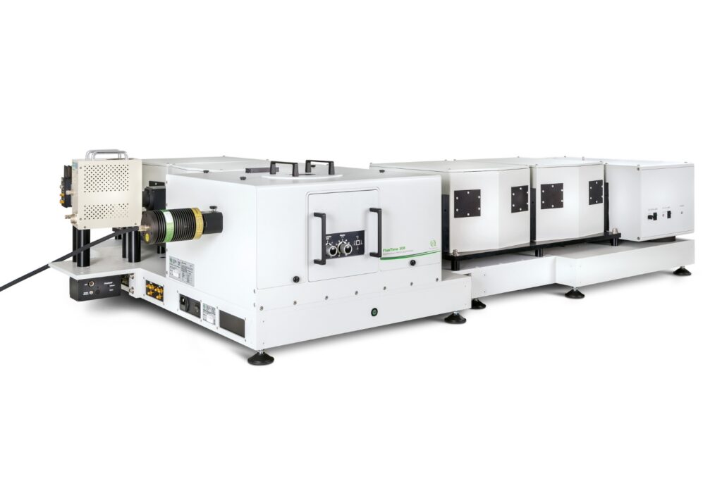
Organic chemistry generally requires toxic and flammable solvents derived from petroleum. These solvents account for ~60% of reaction waste and carry environmental and safety costs.1 Water is a promising alternative, but many organic molecules are neither stable nor soluble in it. Recently, a new approach has emerged: surfactants that can create protected compartments. These compartments are droplets and micelles, where reactions can occur. Yet, these systems are so difficult to study with traditional techniques that their behavior remains poorly understood, slowing down the development of new sustainable and safer reactions.2 Here, we study cross-coupling reactions in water, similar to those used to make pharmaceuticals, agrochemicals, and materials. Fluorescence lifetime imaging microscopy (FLIM) and microspectroscopy (with PicoQuant’s FlexLambda) track the in situ formation of a fluorescent product, revealing that micrometer-scale organic droplets act as both the primary reaction sites and reservoirs for organics. These measurements also disprove a previously hypothesized mechanism, wherein micelles become saturated with product inhibiting catalytic turnover. This study highlights how imaging techniques, and FLIM in particular, can support the rational design of sustainable aqueous organic reactions by revealing the actual nature of colloidal objects, identifying reaction sites, and testing mechanistic hypotheses at the microscale. [1] La Sorella, G., Strukul, G., & Scarso, A., Green Chemistry, 17 (2), 644–683 (2015)
[2] Lipshutz, B. H. Green Chemistry, 26 (2), 739–752 (2024) |
| 13.05 - 14.35 | |
| |
| 14.35 - 15.05 | Samrat Mukhopadhyay, SAS Nagar, Manauli PO, India (Invited Talk) Prying into Biomolecular Condensates Using Single-Molecule FRET and HomoFRETPrying into Biomolecular Condensates Using Single-Molecule FRET and HomoFRET Mukhopadhyay, Samrat Indian Institute of Science Education and Research (IISER) Mohali
Macromolecular phase separation of proteins and nucleic acids into biomolecular condensates is associated with a wide range of cellular functions and deadly neurodegenerative diseases. My laboratory has been actively developing, adapting, and applying a diverse array of emerging methodologies that permit us to delineate the key molecular principles governing macromolecular phase separation. Using site-specific intramolecular single-molecule FRET (Förster resonance energy transfer), we dissected the key molecular events associated with phase separation of the prion-like low-complexity domain of FUS (Fused in Sarcoma), which is linked to the formation of cytoplasmic and nuclear membraneless organelles [1]. Our results showed that a symphony of structural unwinding events converts intramolecular interactions into dynamic crosslinks of multivalent intermolecular contacts, resulting in condensate formation. We also extended our single-molecule FRET studies to investigate condensate-mediated chaperoning of tau that is associated with Alzheimer’s disease [2]. We also demonstrated a unique application of intermolecular homoFRET to study the modulation of emergent material properties of FUS condensates [3]. We further demonstrated that homoFRET imaging can detect and characterize the in-situ formation of cytoplasmic stress granules within mammalian cells. I will also discuss our new results on other RNA-binding proteins involved in stress granule formation [1] A. Joshi, A. Walimbe, A. Avni, S. K. Rai, L. Arora, S. Sarkar & S. Mukhopadhyay. Nature Communications (2023) 14, 7331. [2] S. K. Rai, R. Khanna, A. Sarbahi, A. Joshi & S. Mukhopadhyay Science Advances (2025) 11, eads1241. [3] A. Joshi, A. Walimbe, S. Sarkar, L. Arora, G. Kaur, P. Jhandai, D. Chatterjee, I. Banerjee, S. Mukhopadhyay. Nature Communications (2024) 15, 9215. |
| 15.05 - 15.25 | Georg Krainer, Graz, Austria Single-molecule microfluidics for protein analysis and biomarker sensingSingle-molecule microfluidics for protein analysis and biomarker sensing Georg Krainer University of Graz, Institute of Molecular Biosciences (IMB), Humboldtstraße 50/III, 8010 Graz, Austria
The integration of single-molecule approaches with microfluidics has opened new possibilities for protein analysis and biomarker sensing. By combining the sensitivity of single-molecule fluorescence with the versatility of microfluidic platforms, these methods enable quantitative measurements with rich molecular information. In my talk, I will present our work in this area and introduce recently developed single-molecule microfluidic approaches for protein analysis and biomarker detection. In particular, I will highlight single-molecule microfluidic diffusional sizing (smMDS) [1], which enables quantitative analysis of protein size, interactions, and assembly states in solution at the single-molecule level. I will also present DigitISA [2], a digital microfluidic immunoassay that detects low-abundance protein biomarkers in solution by counting individual immunocomplexes with high sensitivity. Together, these approaches demonstrate the versatility of single-molecule microfluidics for both quantitative protein biophysics and ultrasensitive biomarker detection. [1] Krainer G, Jacquat RPB, Schneider MM, Welsh TJ, Fan J, Peter QAE, Andrzejewska EA, Šneiderienė G, Czekalska MA, Ausserwoeger H, Chai L, Arter WE, Saar KL, Herling TW, Franzmann TM, Kosmoliaptsis V, Alberti S, Hartl F-U, Lee SF, Knowles TPJ. Single-molecule digital sizing of proteins in solution. Nat Commun, 2024, 15, 7740. DOI: https://doi.org/10.1038/s41467-024-50825-9
[2] Krainer G, Saar KL, Arter WE, Welsh TJ, Czekalska MA, Jacquat RPB, Peter Q, Traberg WC, Pujari A, Jayaram AK, Challa P, Taylor CG, van der Linden L-M, Franzmann TM, Owens RM, Alberti S, Klenerman D, Knowles TPJ. Direct digital sensing of protein biomarkers in solution. Nat Commun, 2023, 14, 653. DOI: https://doi.org/10.1038/s41467-023-35792-x |
| 15.25 - 15.45 | Rainer Heintzmann, Jena, Germany Noise-Corrected PCA for Improved Analysis of TCSPC-FLIM DataNoise-Corrected PCA for Improved Analysis of TCSPC-FLIM Data Alix Le Marois3,5, Klaus Suhling4, Rainer Heintzmann1,2 1Leibniz Institute of Photonic Technology, Albert-Einstein Str. 9, 07745 Jena, Germany
2Institute of Physical Chemistry and Abbe Center of Photonics, Friedrich-Schiller-Universität Jena, Helmholtzweg 4, 07743 Jena, Germany
3The Francis Crick Institute, 1 Midland Road, London NW1 1AT
4S6.30, Strand Building, Strand Campus, Strand, London, WC2R 2LS
5Institute Fresnel, Avenue Escadrille Normandie Niemen, 13013 Marseille, France
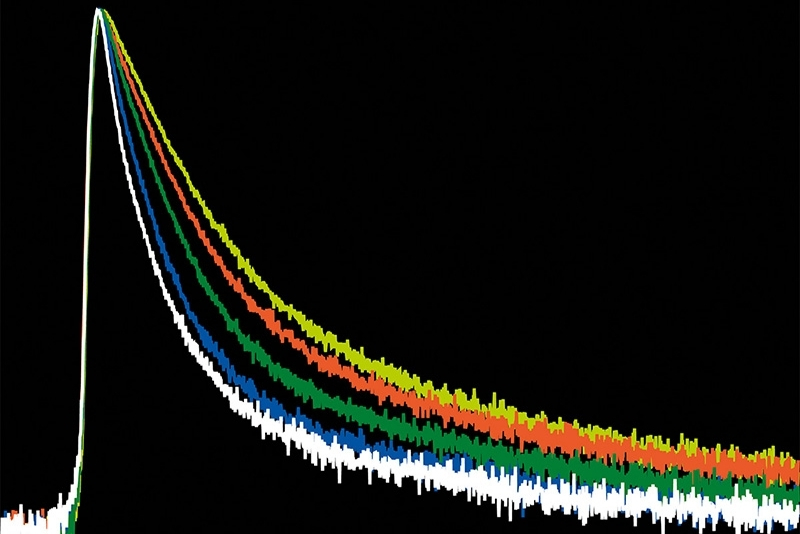
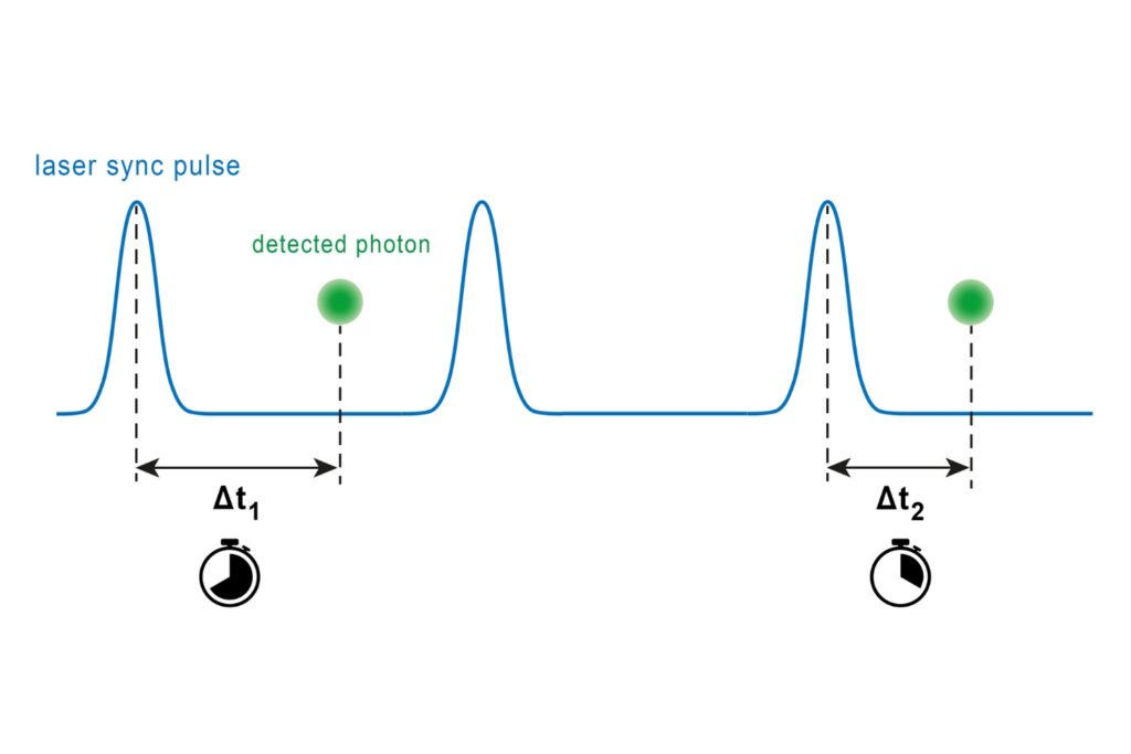
Phasor analysis applied to time-correlated single photon counting (TCSPC) data effectively projects fluorescence decays onto a set of linearly independent subspace coordinates, but in a manner that is sub-optimal with respect to noise. Principal Component Analysis (PCA), in contrast, is designed to identify optimal subspace coordinates that capture the true variance of the underlying data. However, when directly applied to FLIM data, Poisson-distributed photon noise distorts this variance structure and limits PCA performance.
This work introduces a Noise-Corrected PCA (NC-PCA) framework that explicitly accounts for Poisson noise, restoring PCA’s ability to extract meaningful components from photon-limited datasets. The method enables identification of distinct fluorescence decay components without prior knowledge of their number or kinetics and operates effectively at significantly lower photon counts than conventional approaches.
We demonstrate that NC-PCA not only outperforms phasor analysis in resolving microenvironments, but can also serve as a powerful preprocessing step, improving downstream phasor projections. Validation on simulated and experimental data, including membrane imaging in HeLa cells, highlights its robustness and sensitivity to subtle biological variations. A. Le Marois, S. Labouesse, K. Suhling, and R. Heintzmann, Noise-Corrected Principal Component Analysis of fluorescence lifetime imaging data, J. Biophot. 11, 1124-1133, (2016), DOI:10.1002/jbio.201600160
https://github.com/RainerHeintzmann/NCPCA.jl |
| 15.45 - 16.05 | Eli Slenders, Genova, Italy Open-source platform for the analysis of fluorescence fluctuation spectroscopy data with SPAD array detectorsOpen-source platform for the analysis of fluorescence fluctuation spectroscopy data with SPAD array detectors Eli Slenders1, Eleonora Perego2, Sabrina Zappone3, Giuseppe Vicidomini4 1Optical investigation of Dynamical Systems, Istituto Italiano di Tecnologia, Genoa, Italy
2Faculté de biologie et médecine, Université de Lausanne, Lausanne, Switzerland
3Institute of Pharmacy and Molecular Biotechnology, Heidelberg University, Heidelberg, Germany
4Molecular Microscopy and Spectroscopy, Istituto Italiano di Tecnologia, Genoa, Italy
Fluorescence fluctuation spectroscopy (FFS) is a family of techniques for measuring the dynamics of (bio)molecules [1] by analyzing the temporal and/or spatial fluctuations in the fluorescence intensity as fluorophores pass through the detection volume of a confocal microscope. However, by integrating the fluorescence over the sensitive area of the detector, information on the spatial distribution of the photons in the image plane is lost. Recently, the introduction of small single-photon avalanche diode (SPAD) array detectors has opened up a new range of spatiotemporal information, allowing for more detailed analysis of system dynamics [2, 3]. However, the lack of open-source software for analyzing high-dimensional FFS datasets currently limits the adoption of FFS techniques to researchers with programming expertise.
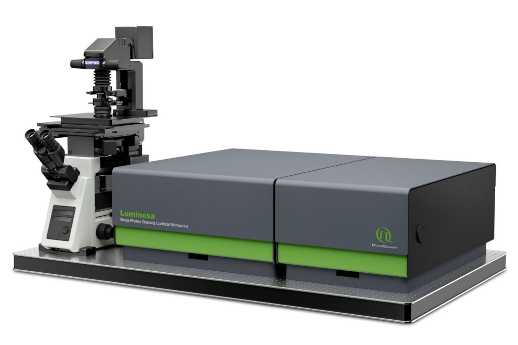
We present BrightEyes-FFS [4], an open-source Python environment for FFS analysis with SPAD array detectors. The environment supports raw data import from BrightEyes-MCS, Genoa Instruments PRISM, and PicoQuant Luminosa. Various algorithms are available for computing correlations, and the resulting curves can be fitted to various models. A graphical user interface and an automated Jupyter Notebook writing tool make the analysis fast and user-friendly. In summary, we believe that BrightEyes-FFS will enable a wider community to study molecular diffusion, flow, and interaction dynamics. [1] D. Magde, W. W. Webb, E. Elson, Phys. Rev. Lett., 29, 1972.
[2] L. Scipioni et al., Nat. Commun., 9, 2018.
[3] E. Slenders et al., Light Sci. Appl., 10, 2021.
[4] E. Slenders et al., BioRxiv., 2026. |
| 16.05 - 16.25 | Jing Liu, West Lafayette, United States Counting fluorescent emitters with a single photon avalanche diode array |
| 16.25 - 16.35 | |
| 16.35 - ... | |