The Quantification Challenge in Living Tissue
Quantifying intracellular ion concentrations in intact biological tissue is not trivial. Intensity-based fluorescence measurements are highly sensitive to dye concentration, excitation fluctuations, and scattering effects. In complex samples, this limits quantitative reliability.
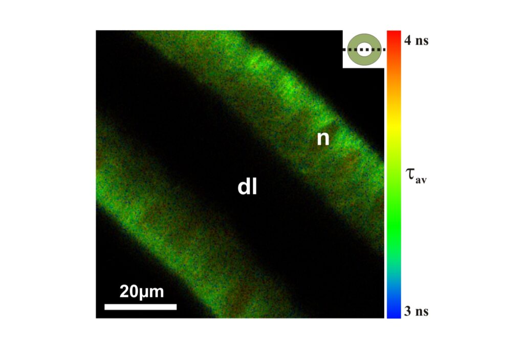
Fluorescence lifetime imaging offers a fundamentally more robust readout. When combined with two-photon excitation, spatial confinement of excitation and reduced photodamage enable measurements under physiological conditions and at greater imaging depth.
Implementing 2P-FLIM on MicroTime 200
The Technical Note describes how two-photon fluorescence lifetime imaging was implemented on MicroTime 200 and experimentally validated.
It outlines:
- verification of true two-photon excitation
- characterization of the instrument response
- integration of TCSPC-based lifetime detection
- in situ calibration strategies for pH- and chloride-sensitive dyes
- quantitative intracellular measurements in living tissue
Rather than discussing 2P-FLIM conceptually, the document presents a complete experimental workflow from optical configuration to calibrated ion concentration maps.
Why This Matters
Quantitative ion imaging requires more than contrast. It requires validated calibration, controlled excitation conditions, and reliable lifetime analysis.
The Technical Note provides the methodological detail necessary to evaluate whether 2P-FLIM is suitable for your biological application.
Download the full Technical Note to explore the complete 2P-FLIM setup, calibration workflow, and quantitative validation data.