Beyond Condensate Morphology
Biomolecular condensates are often first recognized by what can be seen: droplets, domains, or other spatially distinct assemblies that can arise through liquid-liquid phase separation (LLPS).
To understand how condensates form and behave, several molecular questions need to be addressed. Which components accumulate in the dense phase? How are individual molecules organized? How rapidly do they move or change conformation? And how do these molecular properties relate to the behavior of the condensate at larger scales?
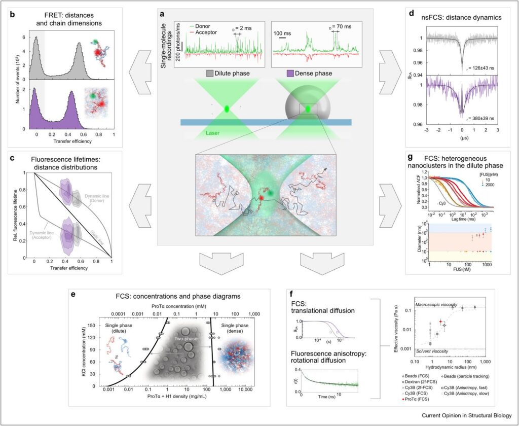
A recent review by Schuler et al., Current Opinion in Structural Biology (2026)1, highlights how single-molecule fluorescence spectroscopy can address these questions. Methods including single-molecule FRET, fluorescence correlation spectroscopy (FCS), fluorescence lifetimes, anisotropy, and nanosecond FCS provide complementary access to molecular structure, concentration, transport, and dynamics across a broad range of length and timescales.
What Is Inside?
The molecular identity of a condensate depends not only on which components are present, but also on how strongly individual species partition into the dense phase.
For a fluorescently labeled component, fluorescence correlation spectroscopy (FCS) can provide quantitative information on molecular concentration. By positioning the confocal observation volume either inside the condensate or in the surrounding dilute phase, the concentration of the labeled species can be compared between both environments.
This makes it possible to move beyond simply observing whether a molecule is present in a condensate and instead quantify how strongly it becomes enriched within the dense phase.
How Are Molecules Organized?
Entering a condensate can alter the molecular state of proteins and nucleic acids.
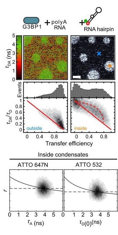
Single-molecule FRET provides access to intramolecular distance distributions and can reveal whether a molecule expands, compacts, unfolds, or shifts between different conformational states. Fluorescence lifetimes provide additional information on these distributions and can help distinguish static heterogeneity from rapid conformational exchange.
This is particularly important for intrinsically disordered proteins and flexible nucleic acids, where a single average structure may not adequately describe the underlying conformational ensemble. No single fluorescence observable captures all of this information. Combining complementary measurements provides a more complete picture of the molecular state within the condensate.
How Do Molecules Move?
Condensates are dynamic, but mobility depends strongly on the length and timescale being considered.
FCS probes translational diffusion, providing information on how molecules move through the dense phase. Time-resolved fluorescence anisotropy addresses rotational mobility, while nanosecond FCS can resolve much faster intramolecular distance fluctuations. These measurements are not interchangeable. Each probes a different aspect of molecular motion.
This distinction becomes particularly important when molecular dynamics are related to material properties such as viscosity. Condensates behave as complex fluids, and the effective viscosity experienced by a molecular probe can depend strongly on its size and on the length scale of the measurement. A condensate can therefore restrict translational motion at larger scales while still allowing rapid rearrangements at the molecular level.
Measuring the Dense and Dilute Phases Separately
Spatial context is another critical part of the measurement.
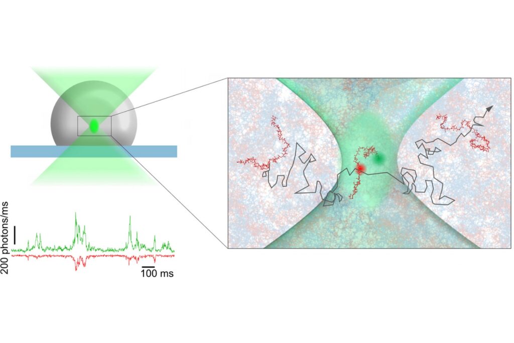
Single-molecule experiments can use very low concentrations of fluorescently labeled molecules within an otherwise predominantly unlabeled sample. The small confocal observation volume can then be positioned selectively within either the dense phase or the surrounding dilute phase.
Individual molecules crossing the observation volume generate photon bursts that can be analyzed for FRET efficiency, fluorescence lifetime, or related molecular properties. The same spatial selectivity enables FCS measurements of concentration and diffusion within defined regions. This connects imaging directly with spectroscopy. Imaging identifies where the measurement should be performed, while precisely positioned single-molecule measurements reveal how molecular behavior changes between environments.
Matching the Measurement to the Question
Functional emergent behavior of condensates depends on molecular interactions across various spatial, temporal, and concentration regimes.
smFRET is particularly sensitive to nanometer-scale structural changes. FCS provides access to molecular concentration and translational diffusion. Fluorescence lifetime and anisotropy add information on molecular environments and mobility. Confocal imaging establishes the spatial context in which these measurements are performed. The measurement strategy therefore has to follow the molecular question rather than relying on a single experimental readout.
Our single photon counting confocal fluorescence microscope Luminosa brings these capabilities together within one confocal platform, including single-molecule FRET, FCS, fluorescence lifetime imaging (FLIM), FLIM-FRET, anisotropy, multichannel single-photon detection, and flexible scanning options. For heterogeneous systems such as biomolecular condensates, the relevant advantage is the ability to combine imaging with quantitative point measurements and to move between complementary fluorescence observables within the same experimental environment.

From Molecular Questions to Condensate Behavior
Understanding biomolecular condensates requires connecting several levels of information.
Concentration and partitioning describe how molecules distribute between phases. Single-molecule FRET and fluorescence lifetimes reveal how molecular conformations change. FCS, anisotropy, and nanosecond FCS probe different forms of molecular motion. Imaging provides the spatial context in which these measurements take place.
These approaches help connect molecular-scale behavior with the properties that emerge at the level of the condensate. The central challenge is therefore not choosing one fluorescence method. It is selecting the observable, spatial scale, and measurement regime that match the molecular question.
1 Reference: B.Schuler, A. Chowdhury, M. T. Ivanović, M. Valdimarsson & N. Galvanetto. Single-molecule fluorescence spectroscopy of biomolecular condensates. Current Opinion in Structural Biology 101, 103354 (2026).
Studying Biomolecular Condensates?
Discuss your experimental requirements with our application specialists and explore how Luminosa can support quantitative fluorescence measurements across structure, dynamics, concentration, and transport.