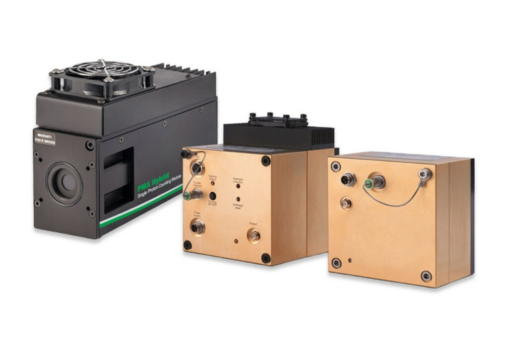
Photon Counting Detectors
High-performance SPAD, PMT and hybrid detectors for reliable photon counting across materials science, life science, quantum optics, and metrology.

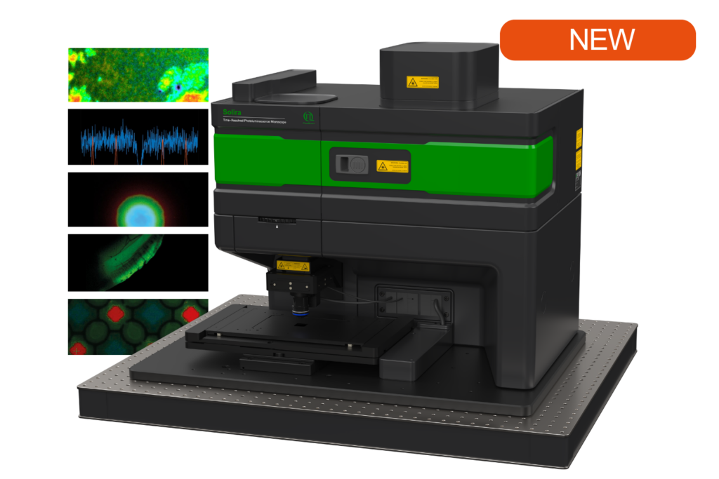
Simplify your materials characterization with one flexible TRPL microscope enabling multiple methods for precise and efficient analysis.

Complete confocal fluorescence microscope that empowers researchers to advance quantitative functional imaging from individual molecules to cells and tissues.

Compact FLIM and FCS upgrade kit that adds advanced functional imaging and correlation analysis to existing laser scanning microscopes.

Designed for flexible, sensitive, and precise steady-state and time-resolved spectroscopy across the UV to NIR range and time scales from picoseconds to milliseconds.

Modular lifetime spectrometer designed for flexible fluorescence and photoluminescence measurements in both materials and life science research.

Add spectral and time-resolved photoluminescence to your setup through flexible microscope–spectrometer coupling options.

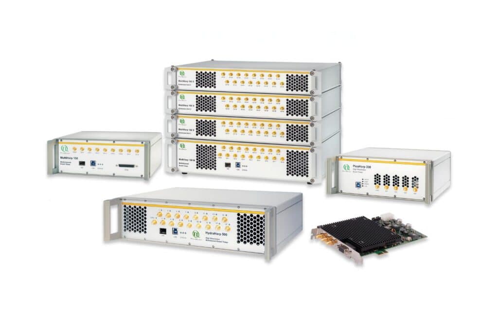
Get the most out of superconducting nanowire detectors in large-scale quantum communication and computing experiments requiring precise multichannel timing.

Boost your time-resolved experiments with a flexible, high-precision time tagging and TCSPC unit for materials science and quantum sensing.

Scale your photonic quantum computing and detector characterization setups while maintaining performance, flexibility, and high data throughput.

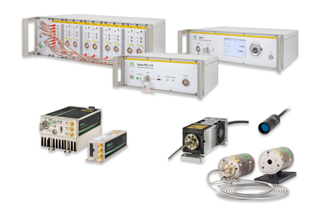
Compact 3-color picosecond laser delivering flexible ns to ms excitation with cost-effective multicolor performance and straightforward operation.

Smart picosecond laser diode heads covering UV-A to NIR, providing the right combination of power, pulse width, and diode type for any time-resolved technique.

VisUV provides clean short pulses and stable timing across key UV and visible wavelengths, including deep UV lines as well as 488 nm and 532 nm.

Enhance your single-photon counting experiments with wide dynamic range and excellent timing precision in the UV and visible even at the highest count rates.

Capture even the weakest signals over large areas with maximum dynamic range and enhanced low-light sensitivity in a compact detector design.

Unlock spatially resolved single-photon detection with a 23-pixel SPAD array, combining low dark counts and precise time tagging for advanced experiments.

Advanced FLIM analysis software for fast, accurate interpretation of lifetime imaging data.

Intuitive, free software solution for real-time, high-precision photon data acquisition, visualization, and initial data analysis.

Advanced software for time-resolved fluorescence acquisition and analysis.

An imaging technique that uses fluorescence lifetimes to generate image contrast.

Investigating how proteins dynamically explore multiple conformational states that control biological function.

Investigating how biomolecules separate into dynamic liquid phases to organize cellular space and regulate biological function.

A time-resolved technique that measures photoluminescence lifetimes to reveal excited-state dynamics in materials.

Studying exciton dynamics, charge carrier processes, and structural properties through optical and time-resolved characterization methods.

Investigating charge-carrier lifetimes and recombination dynamics to enable precise optical characterization of material quality and device performance.

A quantum optical signature revealed by time-resolved photon correlation analysis to identify single-photon emission in materials and nanostructures.

The transmission of information using individual photons, using quantum effects to ensure absolute security.

Quantifying photons per detection event enables direct access to photon-number statistics, providing insight into quantum and statistical properties of light.

An optical technique that analyzes light emission under electrical excitation to reveal electronic properties of electroluminescent materials.

Monitoring environmental signals and trace compounds to understand dynamic changes in natural and engineered environments.

A photon timing technique that measures single-photon arrival times to resolve ultrafast dynamics in fluorescence, materials research, and quantum optics.
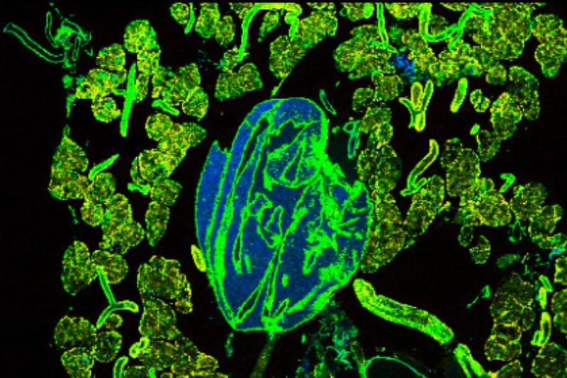
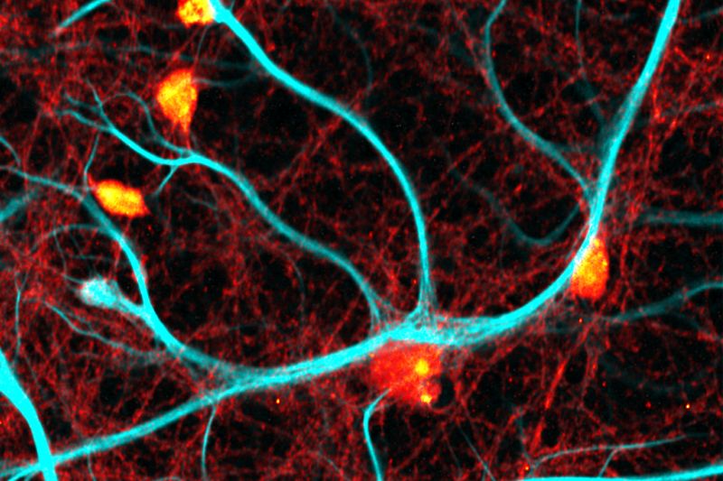
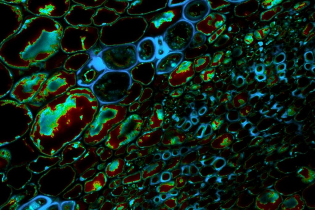
Two-Photon Excitation (TPE) Microscopy (also 2P microscopy, 2PM) is an advanced fluorescence imaging technique used to investigate optically active structures in biological and material samples with highly localized excitation. It enables intrinsic optical sectioning and precise spatial confinement, making it ideal for imaging thick, scattering biological tissues as well as nanostructured and low-dimensional materials. By combining near-infrared excitation with localized fluorescence generation, TPE microscopy achieves intrinsic optical sectioning, reduced photodamage, and greater imaging depth compared to conventional single-photon fluorescence microscopy. It is widely used in life sciences for deep-tissue imaging and in materials science for spatially resolved optical characterization of semiconductors, thin films, and two-dimensional materials.
TPE microscopy relies on the nonlinear absorption of two photons that are absorbed almost simultaneously by a fluorophore or optically active material. This process requires high photon densities and is typically realized using pulsed femtosecond lasers operating in the near-infrared range. Because two-photon absorption occurs only at the focal point of the excitation beam, signal generation is confined to a precisely defined focal volume, providing intrinsic optical sectioning without the need for a confocal pinhole.
Two-photon excitation microscopy generates fluorescence images from intensity signals collected at the focal plane during raster scanning of the sample. Image contrast arises from spatial variations in fluorophore concentration and excitation efficiency. Depending on the experimental setup, TPE data can be further analyzed using advanced techniques such as fluorescence lifetime imaging, spectral unmixing, or correlation analysis. The resulting datasets provide spatially resolved structural and functional insights from thick, scattering biological tissues.
Two-photon excitation microscopy produces spatially resolved intensity maps that reflect the local optical response of the sample. In biological systems, these images reveal structural and functional information through fluorescence contrast. In materials science, TPE data can be integrated with complementary techniques such as time-resolved photoluminescence, second-harmonic generation, or spectral imaging to investigate excitonic behavior, carrier recombination dynamics, and nanoscale material heterogeneity. This multimodal capability enables comprehensive characterization of complex samples across the micro- and nanoscale.
2P microscopy enables highly localized excitation with reduced background fluorescence and improved penetration depth compared to single-photon excitation. The use of near-infrared light reduces scattering and absorption, allowing visualization several hundred micrometers deep within the sample. Intrinsic optical sectioning minimizes background fluorescence and photodamage, making TPE microscopy ideally suited for live-cell and intravital imaging as well as optically sensitive materials.
 Luminosa single photon counting confocal fluorescence microscope designed for quantitative time-resolved and single-molecule imaging.
TPE microscopy requires ultrashort pulsed laser sources delivering high peak powers, typically in the near-infrared spectral range. Lasers that are usually used for this application are Ti:Sa lasers with femtosecond pulse width and a tunable wavelength in the range between 690 nm and 1040 nm. Sensitive photon detection is essential to capture weak signals generated by nonlinear excitation processes. Stable scanning optics and precise synchronization electronics are required for reproducible image acquisition and for combining TPE with time-resolved or nonlinear contrast techniques. Flexible system architectures enable integration of complementary modalities for advanced biological and material characterization.
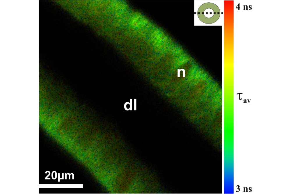
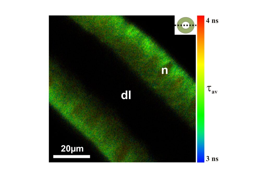
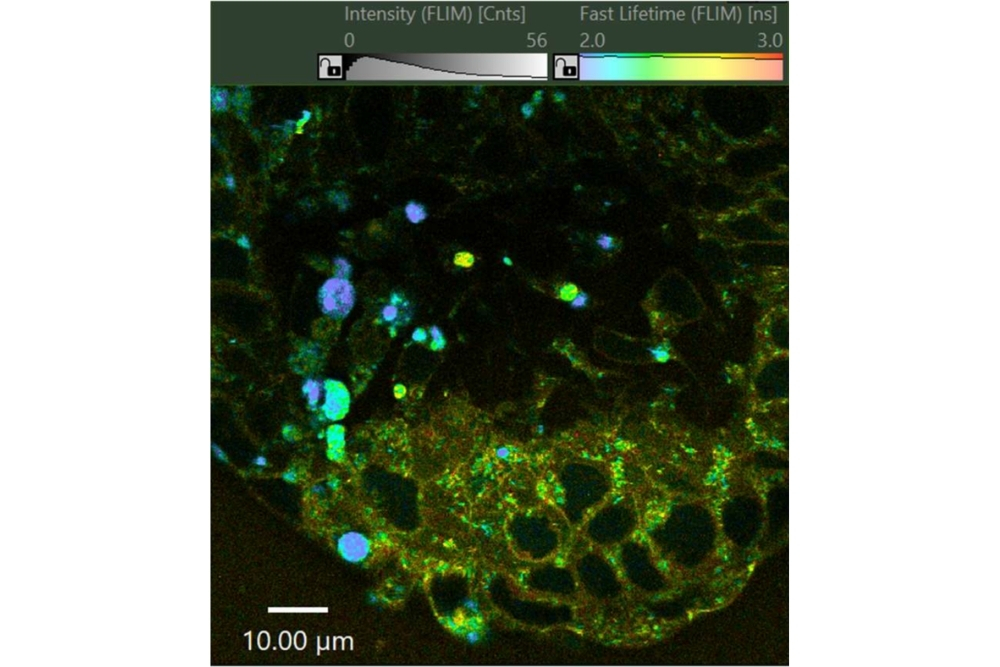
Two-Photon FLIM combines nonlinear excitation with fluorescence lifetime imaging to provide spatially resolved lifetime contrast with intrinsic optical sectioning. By using near-infrared excitation, 2P-FLIM enables deep imaging while reducing photodamage. It is applied to study molecular environments in biological samples and recombination dynamics in optically active materials.
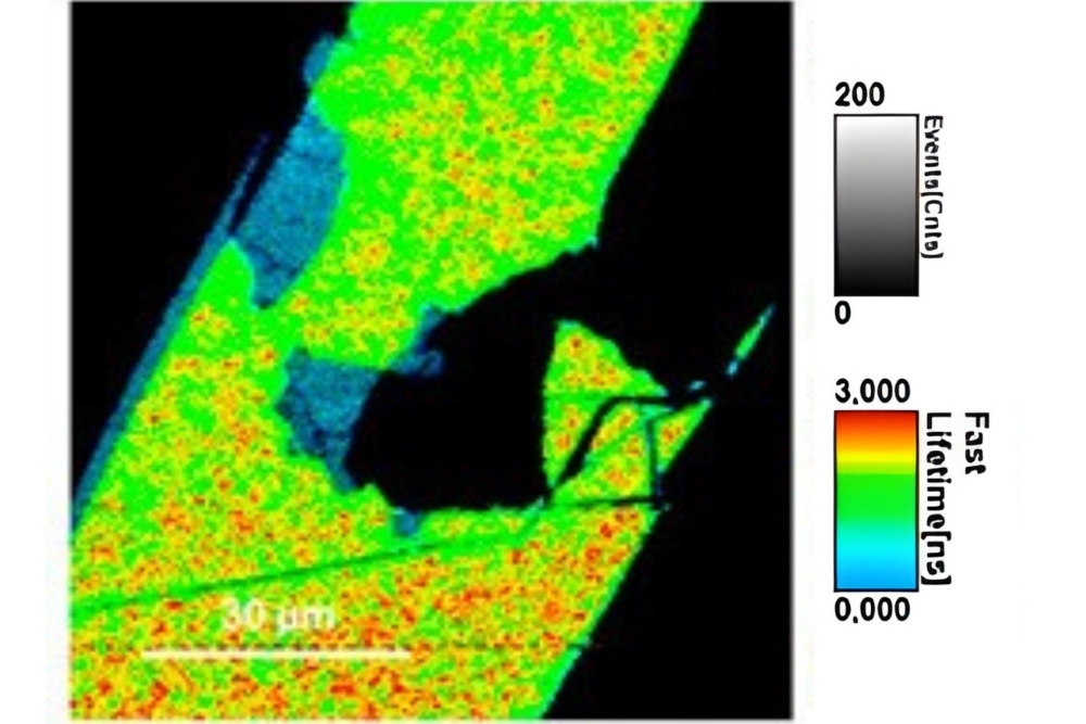
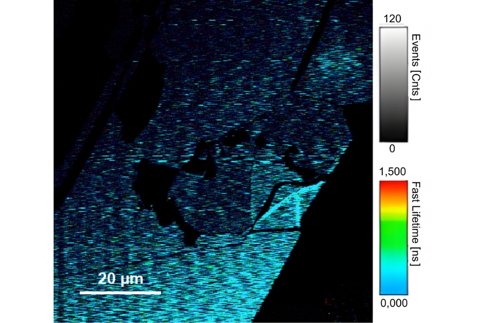
Two-Photon TRPL combines localized nonlinear excitation with time-resolved photoluminescence detection to probe carrier recombination dynamics with high spatial selectivity. It is particularly suited for investigating semiconductors, nanostructures, and 2D materials, enabling depth-resolved analysis of excitonic processes and material heterogeneity.