Photon Counting Detectors
High-performance SPAD, PMT and hybrid detectors for reliable photon counting across materials science, life science, quantum optics, and metrology.

Simplify your materials characterization with one flexible TRPL microscope enabling multiple methods for precise and efficient analysis.

Complete confocal fluorescence microscope that empowers researchers to advance quantitative functional imaging from individual molecules to cells and tissues.

Compact FLIM and FCS upgrade kit that adds advanced functional imaging and correlation analysis to existing laser scanning microscopes.

Designed for flexible, sensitive, and precise steady-state and time-resolved spectroscopy across the UV to NIR range and time scales from picoseconds to milliseconds.

Modular lifetime spectrometer designed for flexible fluorescence and photoluminescence measurements in both materials and life science research.

Add spectral and time-resolved photoluminescence to your setup through flexible microscope–spectrometer coupling options.

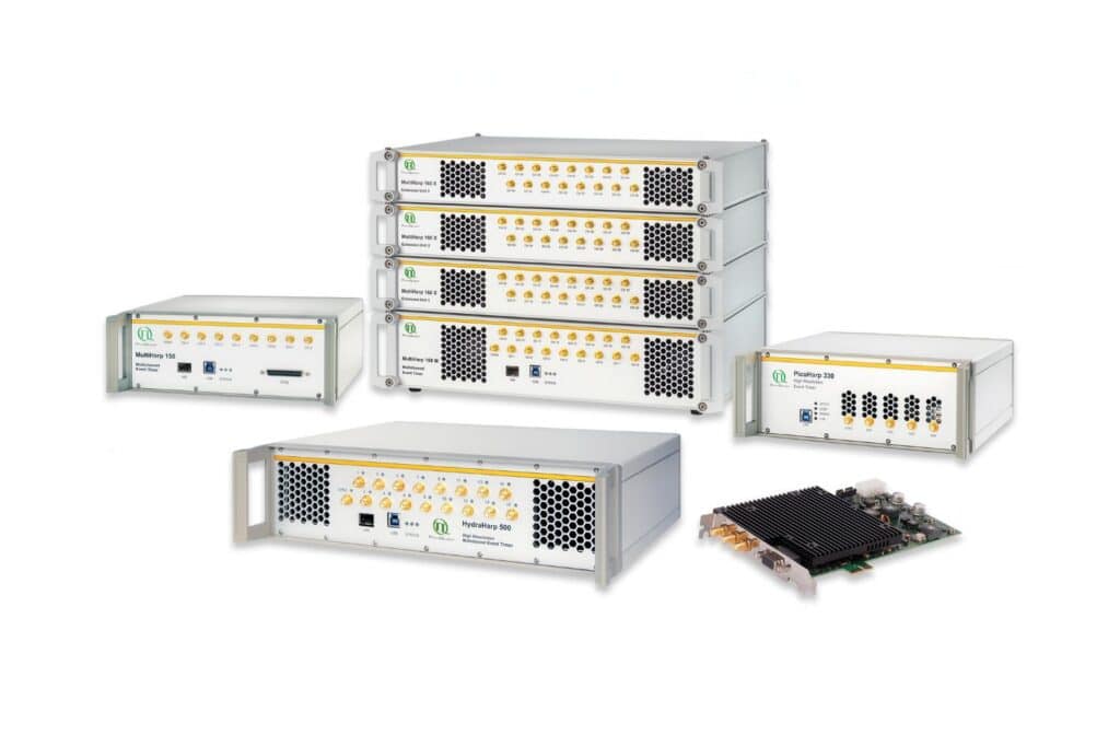
Get the most out of superconducting nanowire detectors in large-scale quantum communication and computing experiments requiring precise multichannel timing.

Boost your time-resolved experiments with a flexible, high-precision time tagging and TCSPC unit for materials science and quantum sensing.

Scale your photonic quantum computing and detector characterization setups while maintaining performance, flexibility, and high data throughput.

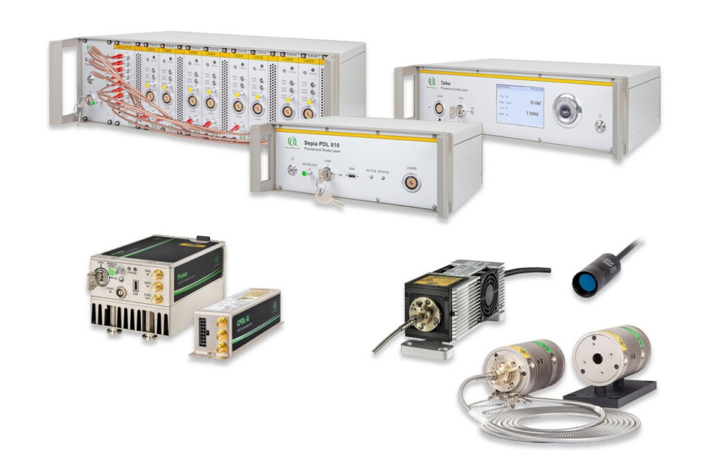
Compact 3-color picosecond laser delivering flexible ns to ms excitation with cost-effective multicolor performance and straightforward operation.

Smart picosecond laser diode heads covering UV-A to NIR, providing the right combination of power, pulse width, and diode type for any time-resolved technique.

VisUV provides clean short pulses and stable timing across key UV and visible wavelengths, including deep UV lines as well as 488 nm and 532 nm.

Enhance your single-photon counting experiments with wide dynamic range and excellent timing precision in the UV and visible even at the highest count rates.

Capture even the weakest signals over large areas with maximum dynamic range and enhanced low-light sensitivity in a compact detector design.

Unlock spatially resolved single-photon detection with a 23-pixel SPAD array, combining low dark counts and precise time tagging for advanced experiments.

Advanced FLIM analysis software for fast, accurate interpretation of lifetime imaging data.

Intuitive, free software solution for real-time, high-precision photon data acquisition, visualization, and initial data analysis.

Advanced software for time-resolved fluorescence acquisition and analysis.

An imaging technique that uses fluorescence lifetimes to generate image contrast.

Investigating how proteins dynamically explore multiple conformational states that control biological function.

Investigating how biomolecules separate into dynamic liquid phases to organize cellular space and regulate biological function.

A time-resolved technique that measures photoluminescence lifetimes to reveal excited-state dynamics in materials.

Studying exciton dynamics, charge carrier processes, and structural properties through optical and time-resolved characterization methods.

Investigating charge-carrier lifetimes and recombination dynamics to enable precise optical characterization of material quality and device performance.

A quantum optical signature revealed by time-resolved photon correlation analysis to identify single-photon emission in materials and nanostructures.

The transmission of information using individual photons, using quantum effects to ensure absolute security.

Quantifying photons per detection event enables direct access to photon-number statistics, providing insight into quantum and statistical properties of light.

An optical technique that analyzes light emission under electrical excitation to reveal electronic properties of electroluminescent materials.

Monitoring environmental signals and trace compounds to understand dynamic changes in natural and engineered environments.

A photon timing technique that measures single-photon arrival times to resolve ultrafast dynamics in fluorescence, materials research, and quantum optics.
Cellular neuroscience focuses on understanding the nervous system at the level of individual neurons, glial cells, and their subcellular components. Rather than describing large-scale brain activity, this field examines how signaling events, molecular interactions, and structural organization within cells give rise to neuronal function. Processes occurring at synapses, membranes, and intracellular compartments are central to cellular neuroscience, providing mechanistic insight into how neural communication is initiated, regulated, and modified at the smallest functional scales.
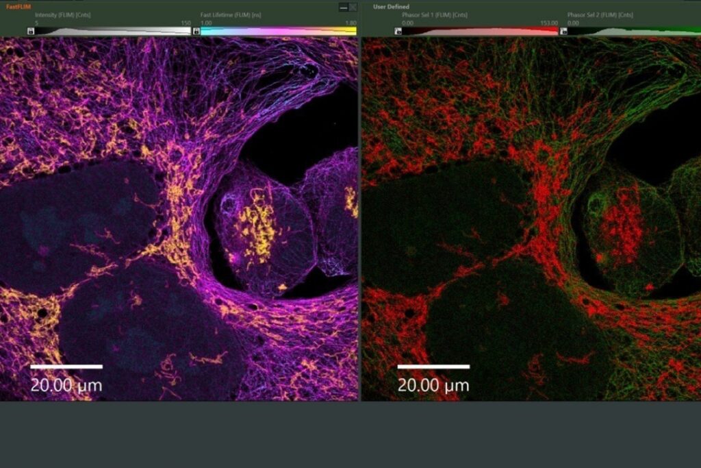
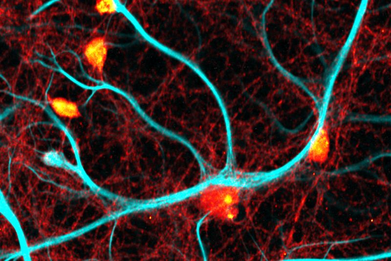
 FLIM image of fixed primary neuronal cells stained for synapses (PSD95), intermediate filaments (GFAP), and mitochondria (TOM20). Sample courtesy of the Rizzoli Group, Department of Neuro- and Sensory Physiology, University of Göttingen Medical Center.
Neuronal function emerges from tightly regulated cellular mechanisms that operate on fast timescales and within highly confined spatial environments. Studying these processes at the cellular level is essential for understanding how synaptic transmission and plasticity support learning, adaptation, and information processing. Disruption of cellular signaling pathways is closely linked to neurological and neurodegenerative diseases, making cellular neuroscience critical for connecting molecular dysfunction to altered neural behavior and pathology.
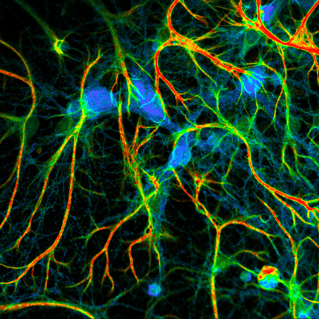
 Presynaptic Bassoon and postsynaptic Homer clusters in fixed neurons visualized by fluorescence lifetime imaging (FLIM), where color encodes lifetime contrast rather than intensity-based staining. Sample courtesy of Rizzoli group, Department of Neuro- and Sensory Physiology, University of Göttingen Medical Center.
Cellular neuroscience investigates a range of dynamic processes that control neural communication. These include neuronal signaling cascades, synaptic transmission, and activity-dependent synaptic plasticity. At a finer scale, protein interactions within synapses, membrane organization, receptor mobility, and metabolic states of neurons and glial cells play decisive roles. These processes determine how signals are transmitted, modulated, and integrated within neural circuits.
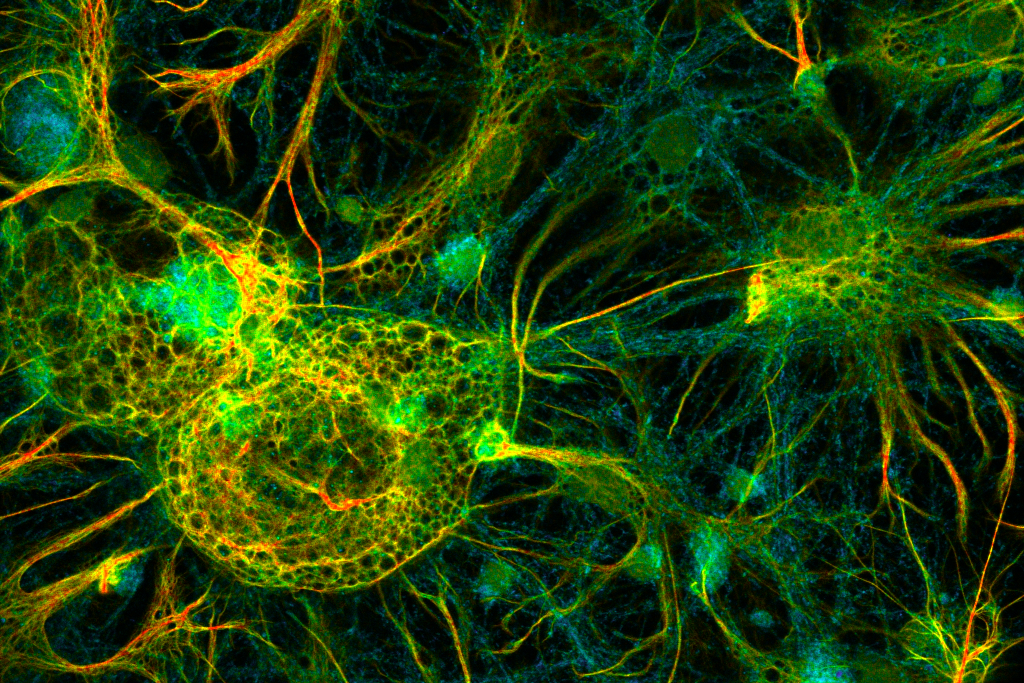
 Fluorescence lifetime imaging (FLIM) of neurons showing cellular morphology and network organization, where color encodes fluorescence lifetime contrast rather than intensity-based staining. Sample courtesy of Rizzoli group, Department of Neuro- and Sensory Physiology, University of Göttingen Medical Center.
Cellular and synaptic processes are highly dynamic and heterogeneous, requiring techniques that provide quantitative insight with high temporal and spatial resolution. Fluorescence-based methods enable noninvasive investigation of living neurons while capturing rapid signaling events and functional changes. Techniques such as fluorescence lifetime imaging microscopy (FLIM), Förster resonance energy transfer (FRET) and fluorescence correlation spectroscopy (FCS) link cellular processes directly to molecular interactions, biochemical states, and neuronal signaling dynamics.
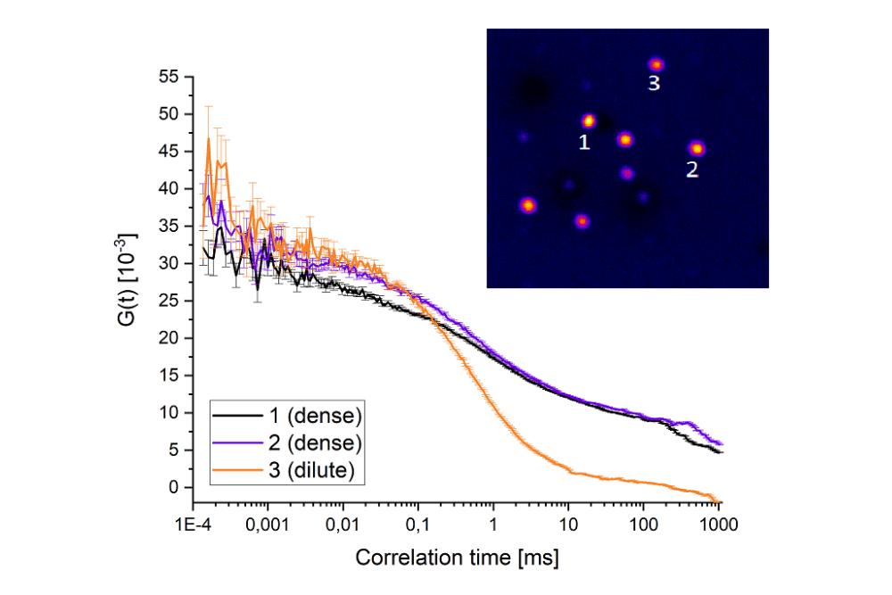
The phase behavior of the neuronal protein synapsin-1 was characterized using fluorescence lifetime imaging (FLIM) and multi-point fluorescence correlation spectroscopy (FCS). Lifetime changes and diffusion analysis showed that synapsin dynamically partitions between condensed and dilute phases within neurons. These liquid-like assemblies contribute to the organization and clustering of synaptic vesicles, illustrating how phase separation mechanisms regulate synaptic architecture and support neuronal signaling at the cellular level.
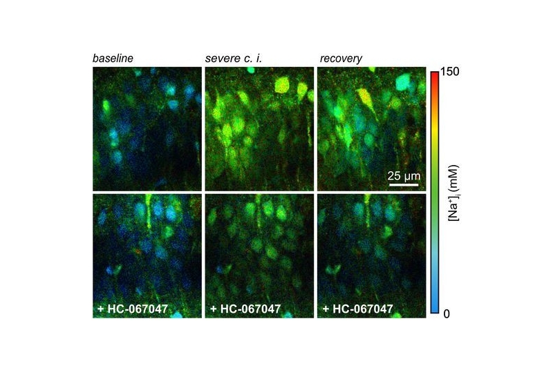
![In situ calibration of ING2. A, ING2-fluorescence of the CA1 region of an organotypic tissue slice. Dashed white lines delineate ROIs drawn around cell bodies from which fluorescence intensity was recorded as shown in B. B, Normalized changes in cellular ING2 fluorescence intensity on changes in [Na+] in one experiment. Gray trace, individual somata; black trace, average. Gray boxes, periods during which FL was recorded as shown in C. C, Same experiment showing color-coded FL images of ING2 taken at different [Na+] with a temporal binning of 15–27 s. Right, Color code. Adapted from Meyer et al. Journal of Neuroscience (2022).](https://www.picoquant.com/wp-content/uploads/flim-sodium-imaging-hippocampal-brain-slice.jpg)
Using fluorescence lifetime imaging, neuronal sodium dynamics were quantitatively monitored in hippocampal brain slices during ischemic conditions. FLIM enabled intensity-independent detection of intracellular ion concentration changes, even under strong tissue movement and volume fluctuations, demonstrating how time-resolved fluorescence provides robust insight into neuronal signaling and synaptic dysfunction in complex neural tissue.
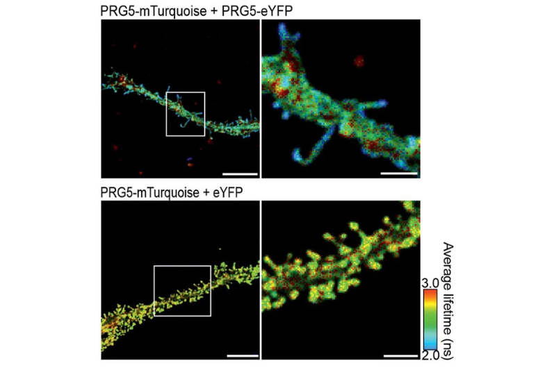
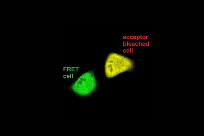
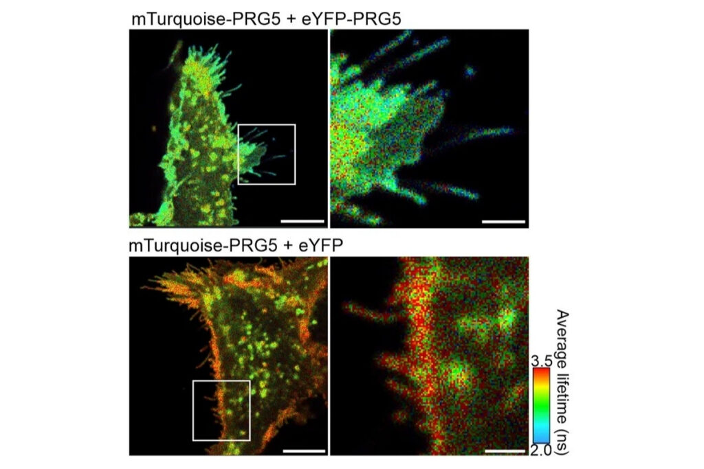
Using fluorescence lifetime imaging combined with FRET, researchers visualized and quantified multimerization of the plasticity-related protein PRG5 at the plasma membrane. FLIM-FRET enabled intensity-independent detection of protein–protein interactions in living cells and primary hippocampal neurons, revealing specific localization of PRG5 multimers at spine-like structures critical for synaptic plasticity.
Using high-speed fluorescence lifetime imaging, rapidFLIMHiRes enabled quantitative monitoring of intracellular Ca²⁺ signaling in living cells following mechanical stimulation. Changes in the fluorescence lifetime of the calcium-sensitive probe Oregon Green BAPTA directly reported transient increases in intracellular calcium concentration, illustrating how fast cellular signaling events relevant to neuronal communication can be captured in real time.