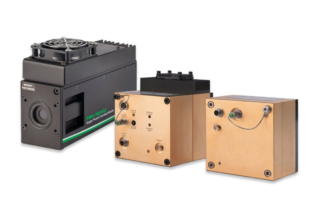
Photon Counting Detectors
High-performance SPAD, PMT and hybrid detectors for reliable photon counting across materials science, life science, quantum optics, and metrology.

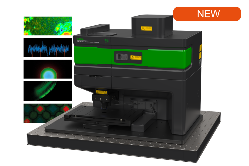
Simplify your materials characterization with one flexible TRPL microscope enabling multiple methods for precise and efficient analysis.

Complete confocal fluorescence microscope that empowers researchers to advance quantitative functional imaging from individual molecules to cells and tissues.

Compact FLIM and FCS upgrade kit that adds advanced functional imaging and correlation analysis to existing laser scanning microscopes.

Designed for flexible, sensitive, and precise steady-state and time-resolved spectroscopy across the UV to NIR range and time scales from picoseconds to milliseconds.

Modular lifetime spectrometer designed for flexible fluorescence and photoluminescence measurements in both materials and life science research.

Add spectral and time-resolved photoluminescence to your setup through flexible microscope–spectrometer coupling options.

Get the most out of superconducting nanowire detectors in large-scale quantum communication and computing experiments requiring precise multichannel timing.

Boost your time-resolved experiments with a flexible, high-precision time tagging and TCSPC unit for materials science and quantum sensing.

Scale your photonic quantum computing and detector characterization setups while maintaining performance, flexibility, and high data throughput.

Compact 3-color picosecond laser delivering flexible ns to ms excitation with cost-effective multicolor performance and straightforward operation.

Smart picosecond laser diode heads covering UV-A to NIR, providing the right combination of power, pulse width, and diode type for any time-resolved technique.

VisUV provides clean short pulses and stable timing across key UV and visible wavelengths, including deep UV lines as well as 488 nm and 532 nm.

Enhance your single-photon counting experiments with wide dynamic range and excellent timing precision in the UV and visible even at the highest count rates.

Capture even the weakest signals over large areas with maximum dynamic range and enhanced low-light sensitivity in a compact detector design.

Unlock spatially resolved single-photon detection with a 23-pixel SPAD array, combining low dark counts and precise time tagging for advanced experiments.

Advanced FLIM analysis software for fast, accurate interpretation of lifetime imaging data.

Intuitive, free software solution for real-time, high-precision photon data acquisition, visualization, and initial data analysis.

Advanced software for time-resolved fluorescence acquisition and analysis.

An imaging technique that uses fluorescence lifetimes to generate image contrast.

Investigating how proteins dynamically explore multiple conformational states that control biological function.

Investigating how biomolecules separate into dynamic liquid phases to organize cellular space and regulate biological function.

A time-resolved technique that measures photoluminescence lifetimes to reveal excited-state dynamics in materials.

Studying exciton dynamics, charge carrier processes, and structural properties through optical and time-resolved characterization methods.

Investigating charge-carrier lifetimes and recombination dynamics to enable precise optical characterization of material quality and device performance.

A quantum optical signature revealed by time-resolved photon correlation analysis to identify single-photon emission in materials and nanostructures.

The transmission of information using individual photons, using quantum effects to ensure absolute security.

Quantifying photons per detection event enables direct access to photon-number statistics, providing insight into quantum and statistical properties of light.

An optical technique that analyzes light emission under electrical excitation to reveal electronic properties of electroluminescent materials.

Monitoring environmental signals and trace compounds to understand dynamic changes in natural and engineered environments.

A photon timing technique that measures single-photon arrival times to resolve ultrafast dynamics in fluorescence, materials research, and quantum optics.
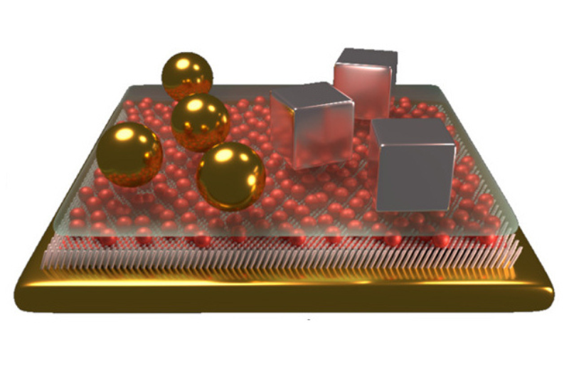
Photoluminescence (PL) is a steady-state optical spectroscopy technique used to study light emission from materials under continuous optical excitation. When photons with sufficient energy promote electrons to higher electronic states, radiative recombination processes emit photons at characteristic energies. The resulting emission spectrum reflects the electronic states and optical transitions of the material. PL is widely applied in materials science, including semiconductor and solar cell research, to evaluate band structure, defect states, and overall material quality.
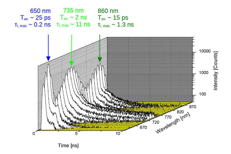
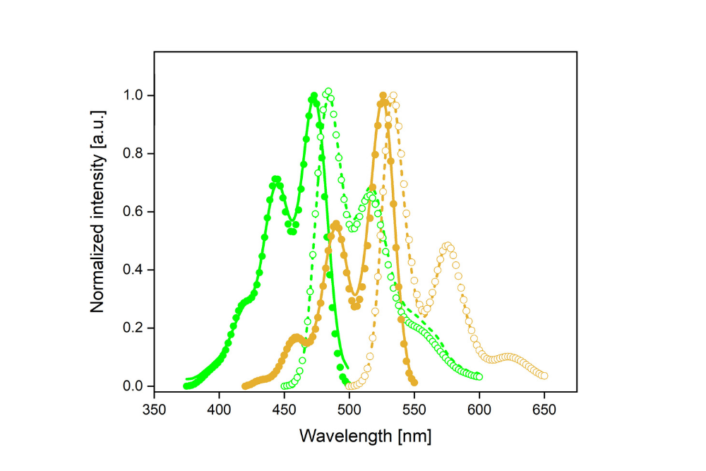
 Example of steady-state emission spectra and time-resolved emission spectra recorded from polymer-based samples, illustrating spectral and temporal photoluminescence analysis.
 In a PL experiment, the sample is illuminated with continuous-wave or pulsed light of energy above its absorption threshold. The absorbed photons excite electrons to higher electronic states, which relax and recombine radiatively, emitting lower-energy photons. The emitted light is spectrally filtered and detected to produce an emission spectrum that reflects the optical transitions within the material. Since PL measurements are performed under steady-state conditions, the technique provides spectral information but does not capture the temporal evolution of emission. To access time-dependent emission dynamics and excited-state lifetimes, photoluminescence can be studied using time-resolved techniques such as Time-Resolved Photoluminescence (TRPL).
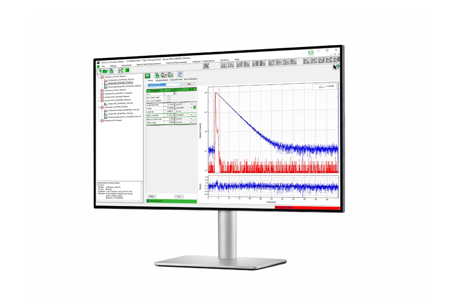 EasyTau 2: spectroscopy control and analysis software
PL data are typically presented as emission spectra showing intensity as a function of wavelength or photon energy. Peak positions provide information on bandgap energies and electronic transitions, while peak shape and linewidth reflect disorder, defect density, and compositional variations. Variations in emission intensity indicate relative radiative efficiency and material quality.
When combined with optical microscopy or spatial scanning, PL measurements can be extended to spatially resolved analysis, commonly referred to as PL imaging or micro-PL. This approach enables mapping of spectral features across a sample to reveal local inhomogeneities, grain boundaries, and defect distributions.
PicoQuant’s EasyTau 2 provides an intuitive software environment for steady-state photoluminescence spectroscopy, enabling rapid visualization, spectral evaluation, and comparison of PL emission data. SymPhoTime 64 extends PL analysis to spatially resolved measurements by combining spectral and imaging workflows for micro-PL and PL imaging experiments within a unified acquisition and analysis platform.
Photoluminescence provides a non-destructive and highly sensitive probe of optically active electronic states in materials. It enables rapid assessment of band structure, defect-related emission, and material uniformity without the need for electrical contacts or elaborate sample preparation. PL is therefore well suited for characterizing semiconductors, nanomaterials, LEDs and other optoelectronic structures, as well as for monitoring material quality during synthesis and device fabrication.
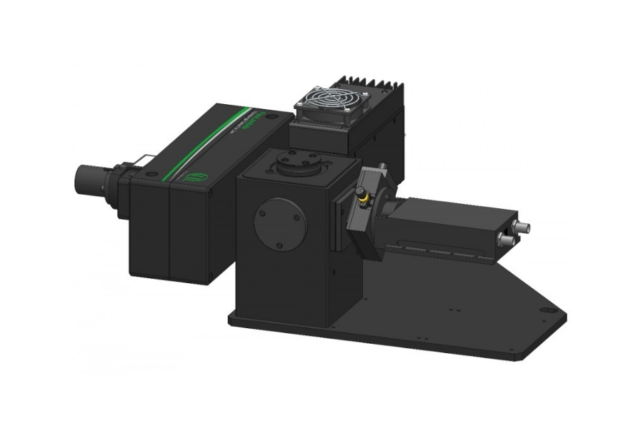
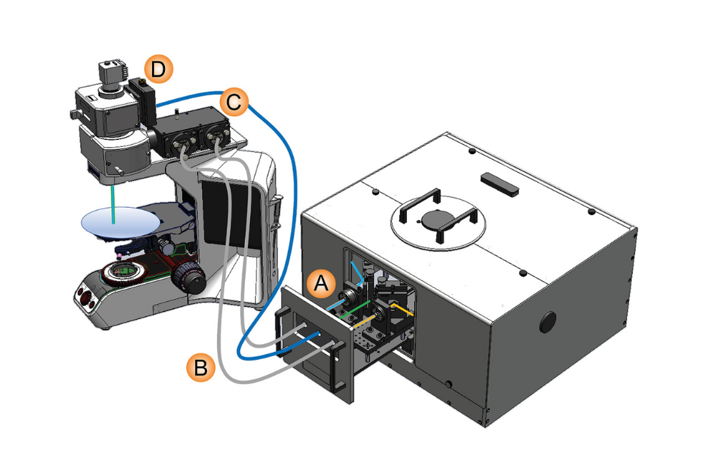 Schematic overview of the FluoMic add-on showing the dedicated sample mounting unit, pre-aligned fiber connections, and fiber in- and out-coupling modules for integration with optical microscopes. Taken from Ermilov et al., Rev. Sci. Instrum. (2020).
Accurate and reproducible PL measurements require a stable optical excitation source, wavelength-selective detection, and high-sensitivity photon detection. Depending on the application, PL can be performed as point spectroscopy or combined with a microscope for spatially resolved measurements.
The following examples demonstrate how steady-state and time-resolved photoluminescence measurements provide spectral and lifetime insight across diverse sample types and spatial scales.
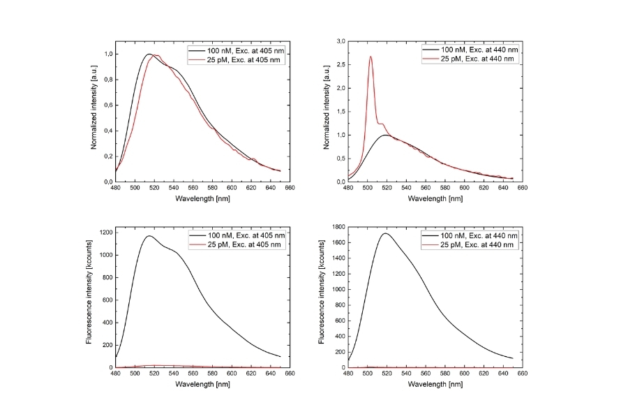
Steady-state emission spectra and fluorescence lifetimes were measured from low-volume Fluorescein samples down to picomolar concentrations. Emission spectra were compared under different excitation wavelengths, and lifetime decays were evaluated using tail-fit and reconvolution analysis. The results demonstrate reliable spectral and temporal characterization even at extremely low signal levels.
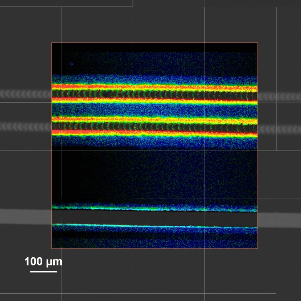
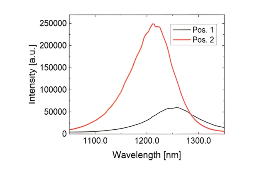
Steady-state spectra, fluorescence decays, and time-resolved emission spectra (TRES) were recorded from well-defined micrometer-sized regions using a spectrometer–microscope assembly. Polymer standards, structured materials, and display pixels were analyzed to correlate spectral, temporal, and spatial information, enabling localized photophysical characterization beyond conventional bulk spectroscopy.
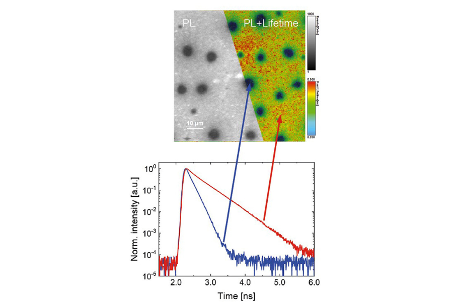
Steady-state emission spectra and time-resolved photoluminescence decays were acquired from defined regions of a CIGS solar cell using variable objective magnifications. Differences in lifetime behavior between measurement spots revealed spatial variations in recombination dynamics, highlighting the importance of localized excitation and detection areas in semiconductor analysis.
A time-resolved spectroscopy technique that records the temporal evolution of photoluminescence emission after pulsed optical excitation. By analyzing the decay of the emitted signal over time, TRPL provides insight into carrier recombination processes and excited-state lifetimes.
An imaging-based extension of TRPL that combines time-resolved detection with spatial scanning or widefield acquisition. TRPL imaging maps photoluminescence lifetimes across a sample, enabling visualization of local variations in recombination dynamics, defects, and material inhomogeneities that are not accessible with spectroscopic measurements alone.