Fluorescence Does Not Measure Structure Directly
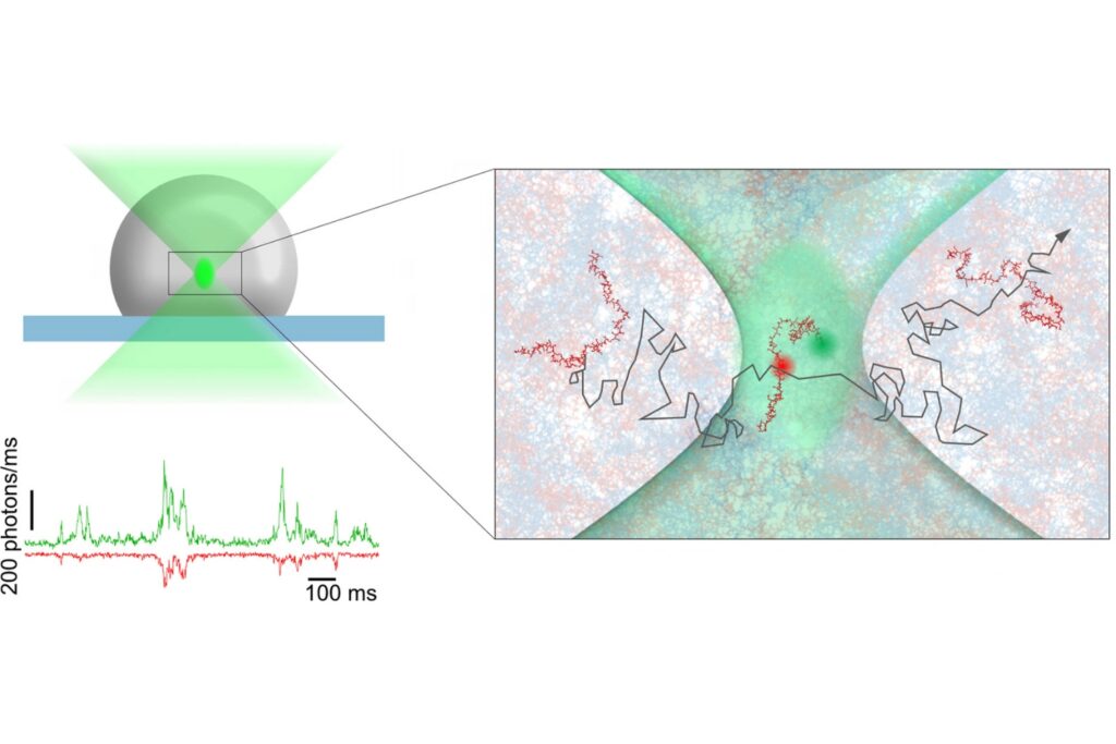
Fluorescence has become an important tool for studying biomolecular conformations, interactions, and dynamics. In a recent review, Peulen et al., Current Opinion in Strucural Biology (2026)1, examine how quantitative fluorescence measurements can be translated into structural and kinetic information about biomolecules. A central challenge is that fluorescence does not report molecular structure directly. Instead, experimentally measured photon signals are converted into intermediate quantities such as FRET efficiencies, distance distributions, rate constants, brightness, or other photophysical parameters. These quantities can then be used to constrain structural or kinetic models.
This distinction is important. The experiment observes the fluorophore, while the scientific question often concerns the biomolecule to which that fluorophore is attached.
Quantitative interpretation therefore depends on a forward model that connects a molecular representation to the experimentally accessible fluorescence signal. The quality of that connection depends strongly on how realistically the fluorophore itself is represented.
The Dye Becomes Part of the Measurement
A fluorescent label is not simply a passive reporter at a fixed position.
Its accessible position, orientation, mobility, interaction with the biomolecular surface, and local environment can all influence the measured fluorescence properties. Different modeling approaches attempt to account for these effects at different levels of complexity.
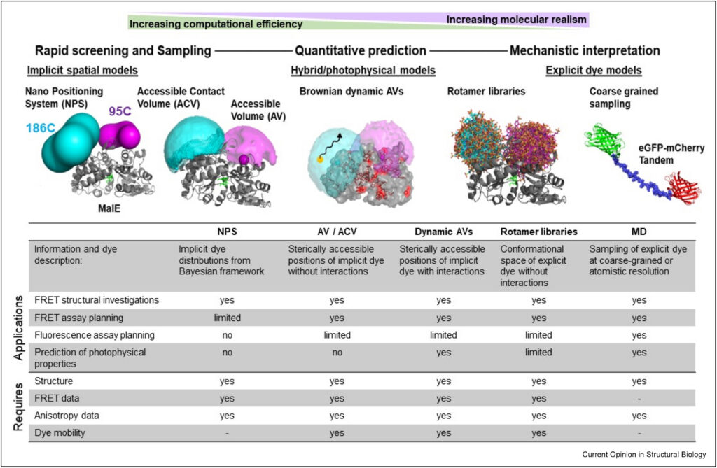
Accessible volume models, for example, estimate the sterically accessible space of a tethered fluorophore. Accessible contact volume models extend this representation by distinguishing mobile and surface-associated dye populations. More detailed approaches incorporate stochastic dynamics, rotamer libraries, coarse-grained representations, or explicit molecular dynamics simulations.
Each approach involves a tradeoff between physical detail and computational efficiency. More importantly, the appropriate model depends on the experimental information available to constrain it. This creates a direct connection between modeling and measurement design.
Why One Observable May Not Be Enough
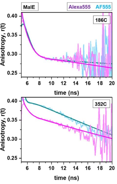
The review highlights fluorescence anisotropy as an example of additional experimental information that can refine a dye model. Restricted rotational motion can indicate interactions between the fluorophore and the molecular surface, helping distinguish mobile from surface-associated dye populations.
Fluorescence lifetime provides another complementary perspective because it is sensitive to the local photophysical environment. Fluorescence lifetime imaging (FLIM) extends lifetime measurements to spatially resolved analysis. Fluorescence correlation spectroscopy (FCS) provides access to fluctuations and diffusion behavior, while single-molecule FRET reports on distance-dependent energy transfer and molecular heterogeneity.
These observables do not necessarily describe the same molecular property. Their value lies in the complementary information they provide about the fluorophore and the molecular system.
A particularly instructive example appears in the review using previously published measurements of AF555 and Alexa Fluor 555 attached to the maltose-binding protein MalE. Although the two cyanine dyes are structurally similar, their fluorescence lifetimes and time-resolved anisotropy decays differ depending on the labeling position. These differences reveal site-specific dye mobility and interactions that would not be captured by a purely steric description of the fluorophore.
For quantitative fluorescence experiments, this has an important consequence: characterizing the dye itself can become part of characterizing the biomolecule.
From Better Models to Better Experimental Constraints
Fluorescence-based structural modeling is an inverse problem. Multiple structural or kinetic models can be compatible with the same experimental information, meaning that measurements rarely determine a unique molecular structure on their own. Increasing model complexity without increasing experimental information therefore has limited value.
The review summarizes this challenge as a balance between the number of independent experimental observations, the molecular detail of the representation, and the effective uncertainty of the model. Sparse spectroscopic data cannot uniquely support arbitrarily detailed molecular structural interpretations.
For experimentalists, this shifts the question. Instead of asking only how precisely one fluorescence parameter can be measured, it becomes equally important to ask which combination of observables provides the information needed for the biological question and the structural model used to interpret it.
Bringing Complementary Fluorescence Measurements Together
Peulen et al. do not evaluate a particular microscope platform. Their analysis nevertheless points toward a practical requirement for quantitative fluorescence research: different experimental observables can provide complementary constraints on fluorophore behavior and molecular models.
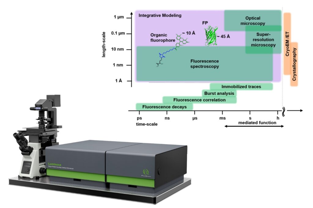
Our fluorescence microscope Luminosa brings several of these measurements together within a single-photon counting confocal platform, including single-molecule FRET, FCS, fluorescence lifetime imaging (FLIM), FLIM-FRET, and fluorescence anisotropy. For single-molecule FRET, pulsed interleaved excitation (PIE) and, for selected wavelengths, alternating laser excitation (ALEX) are available. Up to six point detectors can operate in parallel, while TCSPC acquisition preserves photon timing information for subsequent analysis.

Integrated software supports the transition from photon detection to quantitative analysis. FRETcompass determines correction factors for single-molecule FRET experiments, while InstaFCS provides online correlation analysis. Dedicated workflows are also available for FLIM, FLIM-FRET, and anisotropy imaging. For experiments that require customized or adaptive workflows, LumiPy provides a Python interface to Luminosa, connecting microscope control, online results, and external analysis tools.
Read how LumiPy extends Luminosa with Python workflows >
The value of bringing these capabilities together is not that every experiment requires every fluorescence method. Rather, it gives researchers the flexibility to select complementary measurements according to the information required to characterize the fluorophore, constrain a model, or address a specific biological question.
From Photons to Molecular Structures
Quantitative fluorescence depends not only on increasingly detailed models, but also on experimental observables capable of constraining them. Lifetime, anisotropy, FRET, and correlation measurements provide complementary information about fluorophore behavior and molecular dynamics. Translating observables to structural information requires an integrative approach that takes advantage of complimentary approaches.
An integrated single-photon counting platform such as Luminosa allows researchers to select these measurements according to the information required by the biological system and the model used to interpret it.
1 Reference: T.-O. Peulen, D. Maksutova, T. Cordes, From quantitative modeling of fluorescence experiments on biomolecules to the prediction of spectroscopic dye properties, Current Opinion in Structural Biology 101, 103376 (2026).
Explore Luminosa for Quantitative Fluorescence
Bring single-molecule FRET, FCS, FLIM, FLIM-FRET, and fluorescence anisotropy together within one single-photon counting confocal platform.