Photon Counting Detectors
High-performance SPAD, PMT and hybrid detectors for reliable photon counting across materials science, life science, quantum optics, and metrology.

Simplify your materials characterization with one flexible TRPL microscope enabling multiple methods for precise and efficient analysis.

Complete confocal fluorescence microscope that empowers researchers to advance quantitative functional imaging from individual molecules to cells and tissues.

Compact FLIM and FCS upgrade kit that adds advanced functional imaging and correlation analysis to existing laser scanning microscopes.

Designed for flexible, sensitive, and precise steady-state and time-resolved spectroscopy across the UV to NIR range and time scales from picoseconds to milliseconds.

Modular lifetime spectrometer designed for flexible fluorescence and photoluminescence measurements in both materials and life science research.

Add spectral and time-resolved photoluminescence to your setup through flexible microscope–spectrometer coupling options.

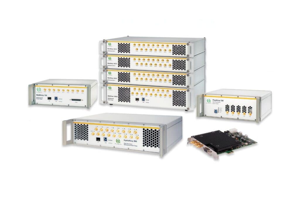
Get the most out of superconducting nanowire detectors in large-scale quantum communication and computing experiments requiring precise multichannel timing.

Boost your time-resolved experiments with a flexible, high-precision time tagging and TCSPC unit for materials science and quantum sensing.

Scale your photonic quantum computing and detector characterization setups while maintaining performance, flexibility, and high data throughput.

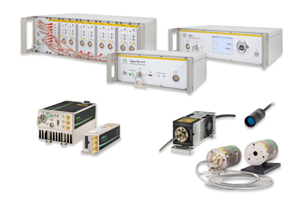
Compact 3-color picosecond laser delivering flexible ns to ms excitation with cost-effective multicolor performance and straightforward operation.

Smart picosecond laser diode heads covering UV-A to NIR, providing the right combination of power, pulse width, and diode type for any time-resolved technique.

VisUV provides clean short pulses and stable timing across key UV and visible wavelengths, including deep UV lines as well as 488 nm and 532 nm.

Enhance your single-photon counting experiments with wide dynamic range and excellent timing precision in the UV and visible even at the highest count rates.

Capture even the weakest signals over large areas with maximum dynamic range and enhanced low-light sensitivity in a compact detector design.

Unlock spatially resolved single-photon detection with a 23-pixel SPAD array, combining low dark counts and precise time tagging for advanced experiments.

Advanced FLIM analysis software for fast, accurate interpretation of lifetime imaging data.

Intuitive, free software solution for real-time, high-precision photon data acquisition, visualization, and initial data analysis.

Advanced software for time-resolved fluorescence acquisition and analysis.

An imaging technique that uses fluorescence lifetimes to generate image contrast.

Investigating how proteins dynamically explore multiple conformational states that control biological function.

Investigating how biomolecules separate into dynamic liquid phases to organize cellular space and regulate biological function.

A time-resolved technique that measures photoluminescence lifetimes to reveal excited-state dynamics in materials.

Studying exciton dynamics, charge carrier processes, and structural properties through optical and time-resolved characterization methods.

Investigating charge-carrier lifetimes and recombination dynamics to enable precise optical characterization of material quality and device performance.

A quantum optical signature revealed by time-resolved photon correlation analysis to identify single-photon emission in materials and nanostructures.

The transmission of information using individual photons, using quantum effects to ensure absolute security.

Quantifying photons per detection event enables direct access to photon-number statistics, providing insight into quantum and statistical properties of light.

An optical technique that analyzes light emission under electrical excitation to reveal electronic properties of electroluminescent materials.

Monitoring environmental signals and trace compounds to understand dynamic changes in natural and engineered environments.

A photon timing technique that measures single-photon arrival times to resolve ultrafast dynamics in fluorescence, materials research, and quantum optics.
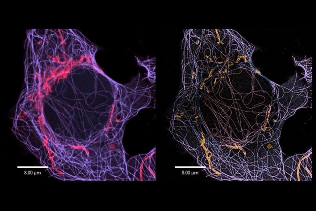
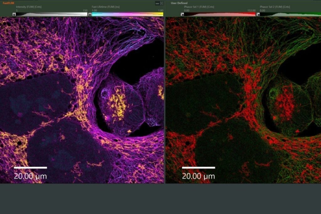
Image Scanning Microscopy (ISM) is an advanced confocal imaging technique that enhances spatial resolution and contrast beyond conventional confocal microscopy. Instead of using a single-point detector, ISM employs a detector array to record spatially resolved fluorescence at each scan position. Through computational pixel reassignment, detected photons are mapped to their most probable point of origin, thereby improving image resolution while preserving optical sectioning. ISM is compatible with standard fluorescent probes and integrates seamlessly with functional imaging modalities such as fluorescence lifetime imaging.
In Image Scanning Microscopy (ISM), fluorescence is detected using a multi-element detector array instead of a single pinhole detector. Each detector element effectively functions as a miniature confocal pinhole, maintaining strong optical sectioning. During image reconstruction, detected signals are computationally shifted toward their true emission origin through pixel reassignment, which enhances spatial resolution beyond conventional confocal imaging. Further improvements in contrast and detail can be achieved with focus-variant ISM and optional deconvolution algorithms.
ISM data comprise spatially resolved intensity maps reconstructed from detector array signals via pixel reassignment. ISM data can be analyzed with standard image processing tools or combined with fluorescence lifetime imaging for ISM-FLIM. In ISM-FLIM, spatial resolution enhancement is directly combined with lifetime-based contrast, enabling improved discrimination of fluorophores and functional states within the same dataset. Additional resolution enhancement may be achieved through computational sectioning algorithms.
Computational sectioning further improves optical sectioning and image contrast by selectively suppressing out-of-focus signal contributions using detector array information. Unlike pixel reassignment, which primarily enhances lateral resolution, computational sectioning increases axial discrimination and reduces background. This results in clearer visualization of fine structural details, particularly in thick or weakly scattering samples.
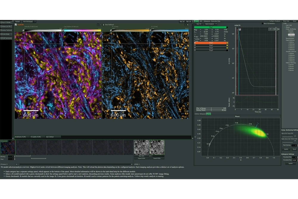 NovaISM software interface for Image Scanning Microscopy data reconstruction and analysis, integrating pixel reassignment, computational sectioning, deconvolution, and fluorescence lifetime evaluation within a unified workflow.
NovaISM is dedicated software for the reconstruction and analysis of Image Scanning Microscopy data. It integrates pixel reassignment, computational sectioning, and deconvolution to enhance spatial resolution and optical sectioning in a unified workflow.
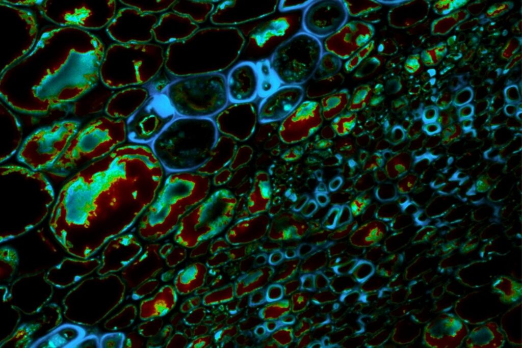
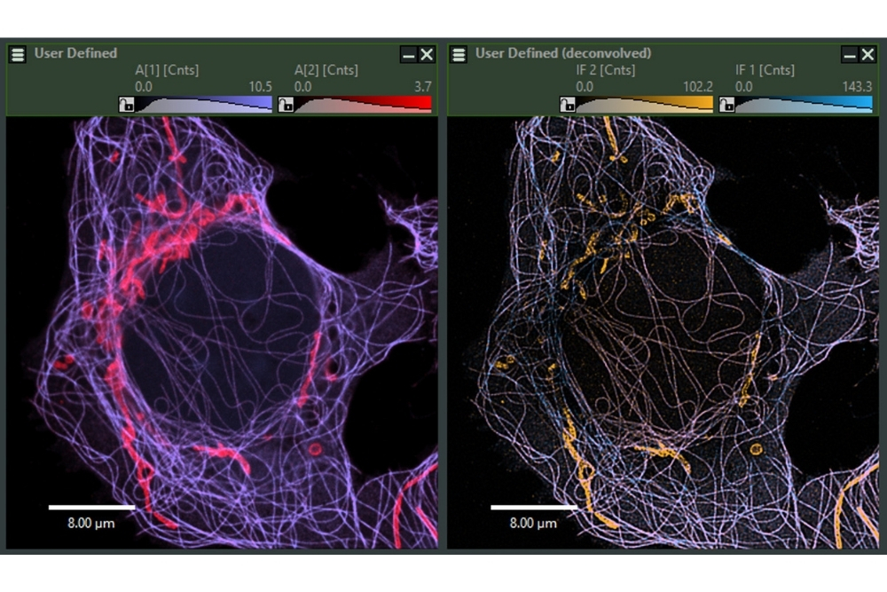 ISM-FLIM species separation in U2OS cells labeled for mitochondria (Tom20, Alexa Fluor 532) and microtubules (α-Tubulin, Alexa Fluor 555). Left: separation after ISM reconstruction. Right: enhanced separation and contrast following computational sectioning and deconvolution.
ISM provides improved spatial resolution and contrast while maintaining the optical sectioning capability and system stability characteristic of confocal microscopy. Because it does not require specialized fluorophores or high illumination intensities, ISM is well suited for sensitive biological samples. The method enables clearer visualization of fine structural details and can be combined with functional contrast mechanisms such as fluorescence lifetime imaging. ISM therefore offers an efficient and accessible approach to resolution-enhanced confocal imaging.
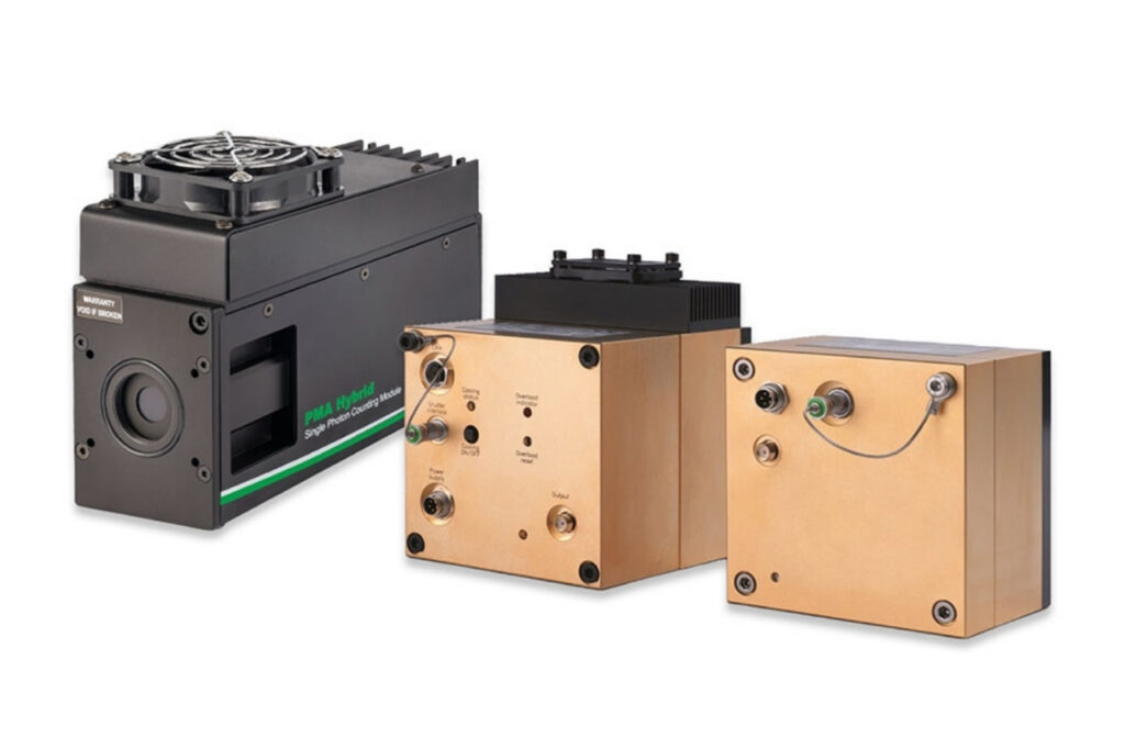
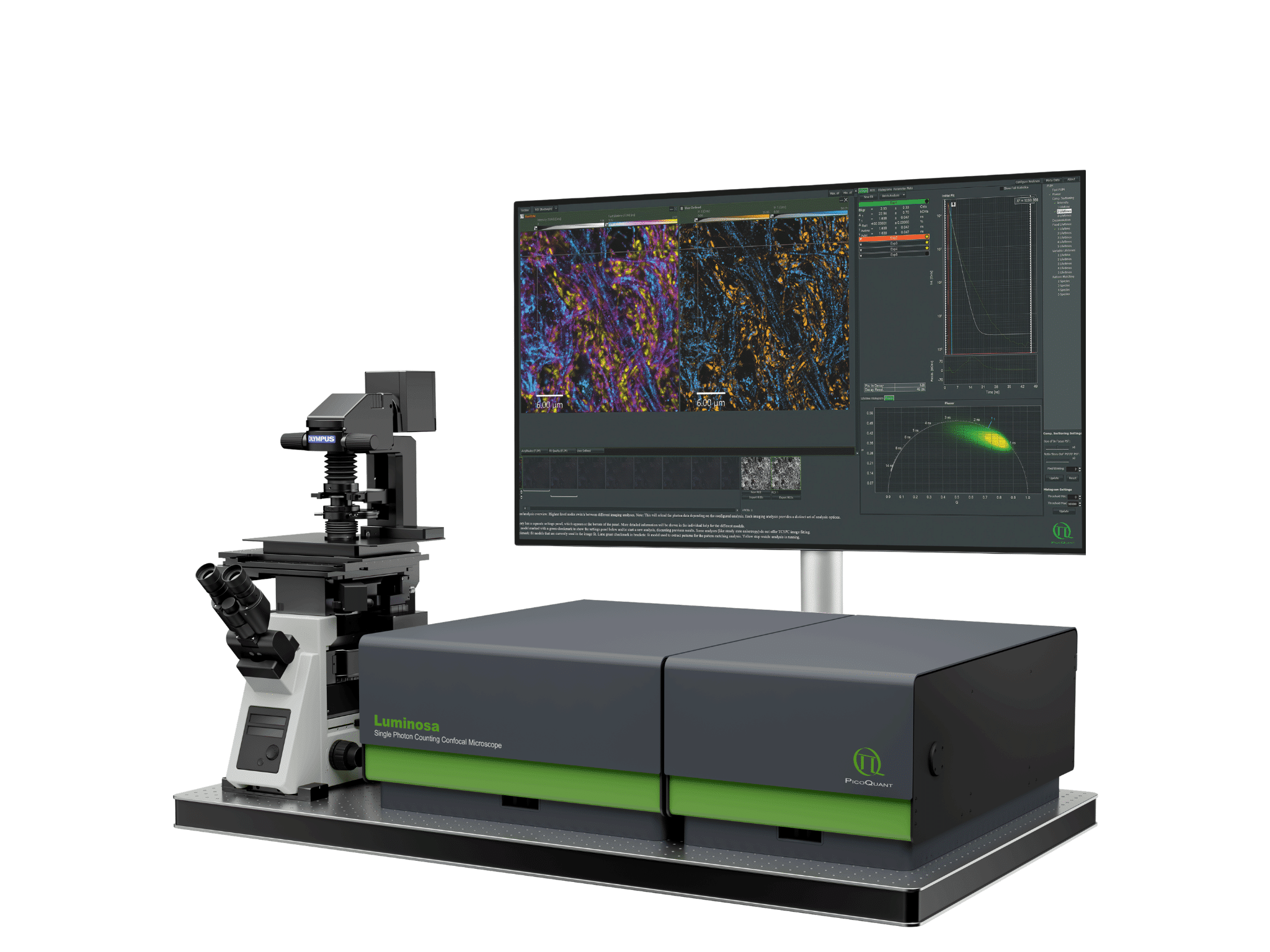 Luminosa confocal microscope combined with NovaISM enables advanced ISM-FLIM imaging and analysis workflows.
Robust Image Scanning Microscopy measurements require confocal laser scanning combined with spatially resolved photon detection and precise data acquisition. Key instrumentation requirements include:
Together, these components provide resolution-enhanced confocal imaging while maintaining optical sectioning capability and quantitative fluorescence contrast.