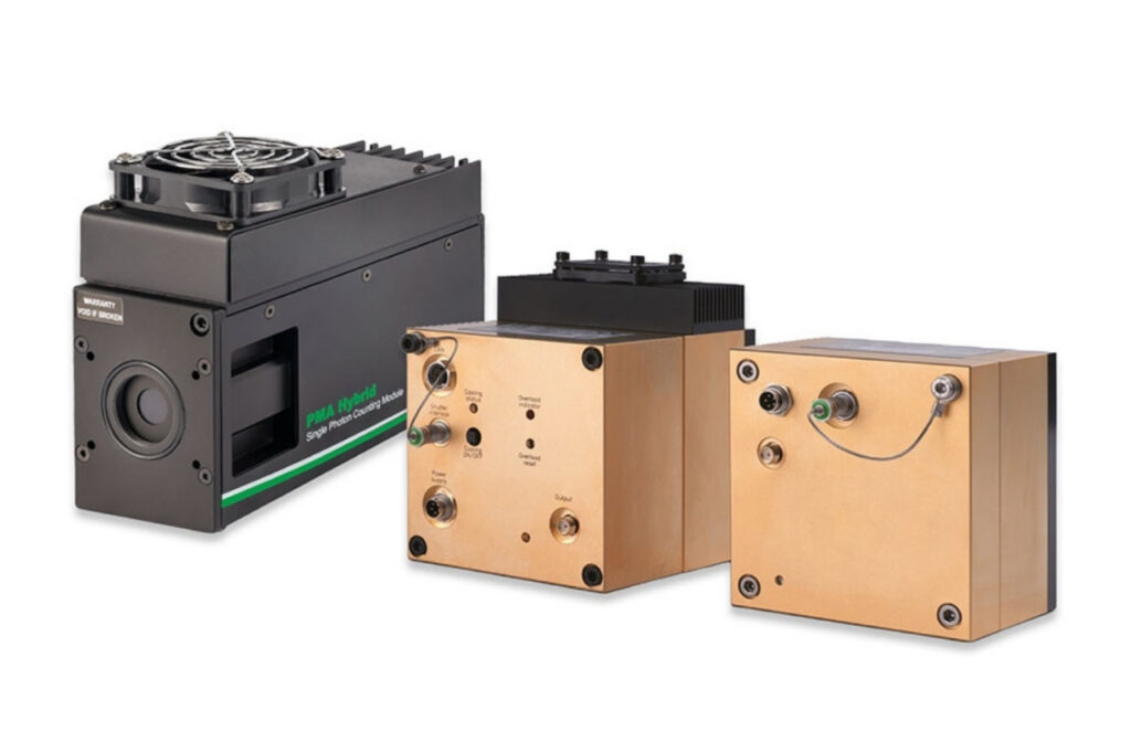
Photon Counting Detectors
High-performance SPAD, PMT and hybrid detectors for reliable photon counting across materials science, life science, quantum optics, and metrology.

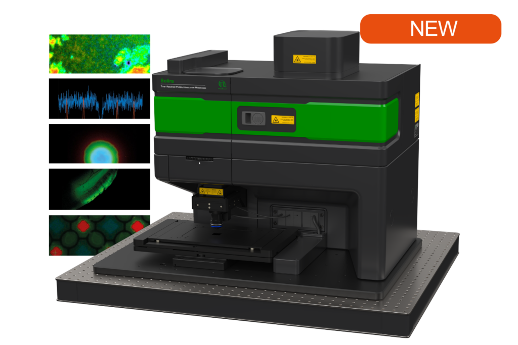
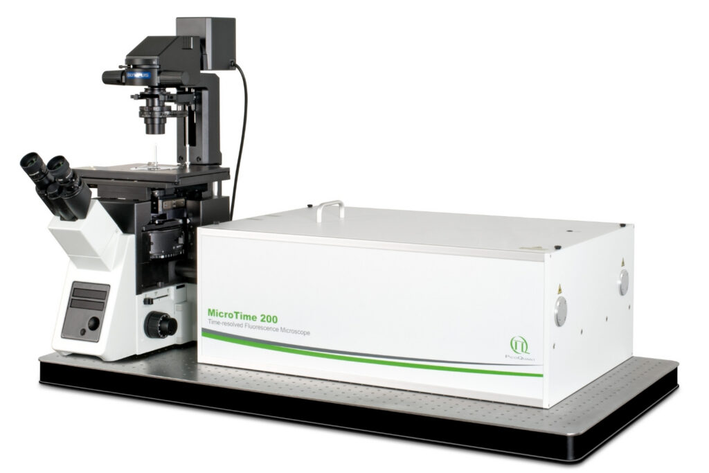
Simplify your materials characterization with one flexible TRPL microscope enabling multiple methods for precise and efficient analysis.

Complete confocal fluorescence microscope that empowers researchers to advance quantitative functional imaging from individual molecules to cells and tissues.

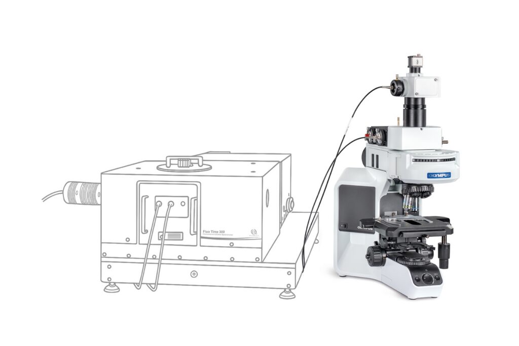
Compact FLIM and FCS upgrade kit that adds advanced functional imaging and correlation analysis to existing laser scanning microscopes.

Designed for flexible, sensitive, and precise steady-state and time-resolved spectroscopy across the UV to NIR range and time scales from picoseconds to milliseconds.

Modular lifetime spectrometer designed for flexible fluorescence and photoluminescence measurements in both materials and life science research.

Add spectral and time-resolved photoluminescence to your setup through flexible microscope–spectrometer coupling options.

Get the most out of superconducting nanowire detectors in large-scale quantum communication and computing experiments requiring precise multichannel timing.

Boost your time-resolved experiments with a flexible, high-precision time tagging and TCSPC unit for materials science and quantum sensing.

Scale your photonic quantum computing and detector characterization setups while maintaining performance, flexibility, and high data throughput.

Compact 3-color picosecond laser delivering flexible ns to ms excitation with cost-effective multicolor performance and straightforward operation.

Smart picosecond laser diode heads covering UV-A to NIR, providing the right combination of power, pulse width, and diode type for any time-resolved technique.

VisUV provides clean short pulses and stable timing across key UV and visible wavelengths, including deep UV lines as well as 488 nm and 532 nm.

Enhance your single-photon counting experiments with wide dynamic range and excellent timing precision in the UV and visible even at the highest count rates.

Capture even the weakest signals over large areas with maximum dynamic range and enhanced low-light sensitivity in a compact detector design.

Unlock spatially resolved single-photon detection with a 23-pixel SPAD array, combining low dark counts and precise time tagging for advanced experiments.

Advanced FLIM analysis software for fast, accurate interpretation of lifetime imaging data.

Intuitive, free software solution for real-time, high-precision photon data acquisition, visualization, and initial data analysis.

Advanced software for time-resolved fluorescence acquisition and analysis.

An imaging technique that uses fluorescence lifetimes to generate image contrast.

Investigating how proteins dynamically explore multiple conformational states that control biological function.

Investigating how biomolecules separate into dynamic liquid phases to organize cellular space and regulate biological function.

A time-resolved technique that measures photoluminescence lifetimes to reveal excited-state dynamics in materials.

Studying exciton dynamics, charge carrier processes, and structural properties through optical and time-resolved characterization methods.

Investigating charge-carrier lifetimes and recombination dynamics to enable precise optical characterization of material quality and device performance.

A quantum optical signature revealed by time-resolved photon correlation analysis to identify single-photon emission in materials and nanostructures.

The transmission of information using individual photons, using quantum effects to ensure absolute security.

Quantifying photons per detection event enables direct access to photon-number statistics, providing insight into quantum and statistical properties of light.

An optical technique that analyzes light emission under electrical excitation to reveal electronic properties of electroluminescent materials.

Monitoring environmental signals and trace compounds to understand dynamic changes in natural and engineered environments.

A photon timing technique that measures single-photon arrival times to resolve ultrafast dynamics in fluorescence, materials research, and quantum optics.
Time-resolved photoluminescence (TRPL) imaging is a spatially resolved, non-invasive extension of the TRPL technique that measures photoluminescence (PL) decay dynamics across a sample. By determining PL lifetimes at each pixel, it reveals local variations in recombination dynamics, defect distributions, carrier diffusion, and material quality. Compared to steady-state photoluminescence, TRPL imaging combines temporal and spatial resolution to probe excited-state processes in detail. The technique is widely applied in materials science to investigate semiconductors, solar cells, light-emitting devices, nanostructures, and two-dimensional materials, supporting fundamental studies and device optimization.
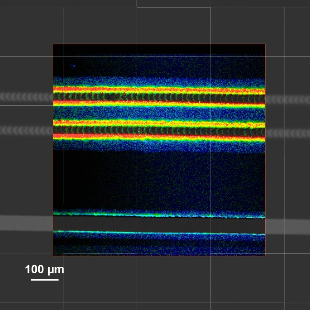
 TRPL imaging of CdTe wafers. Left: Intensity and lifetime images of a CdTe wafer before (a, d) and after thermal activation (b, e). Right: Statistical distribution of intensities (c) and lifetimes (e, f) before (blue) and after (green) thermal activation.
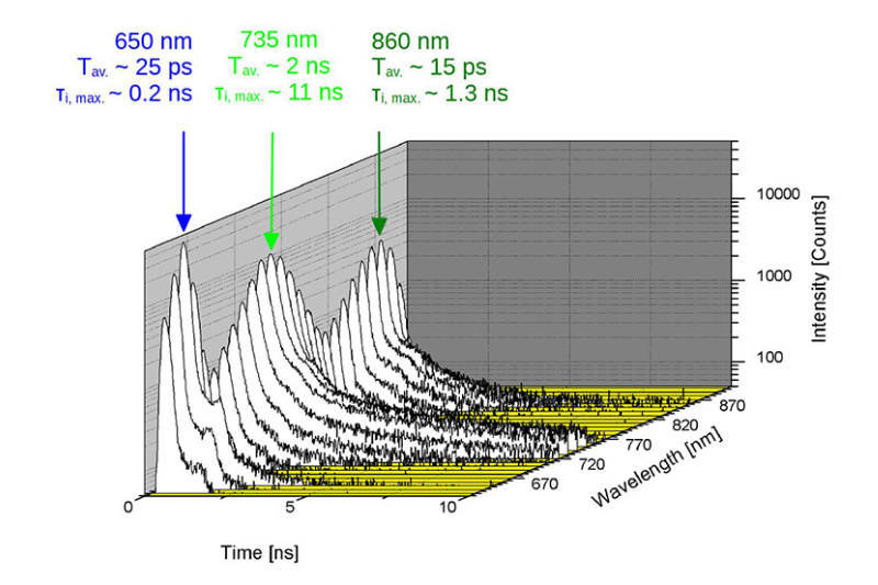
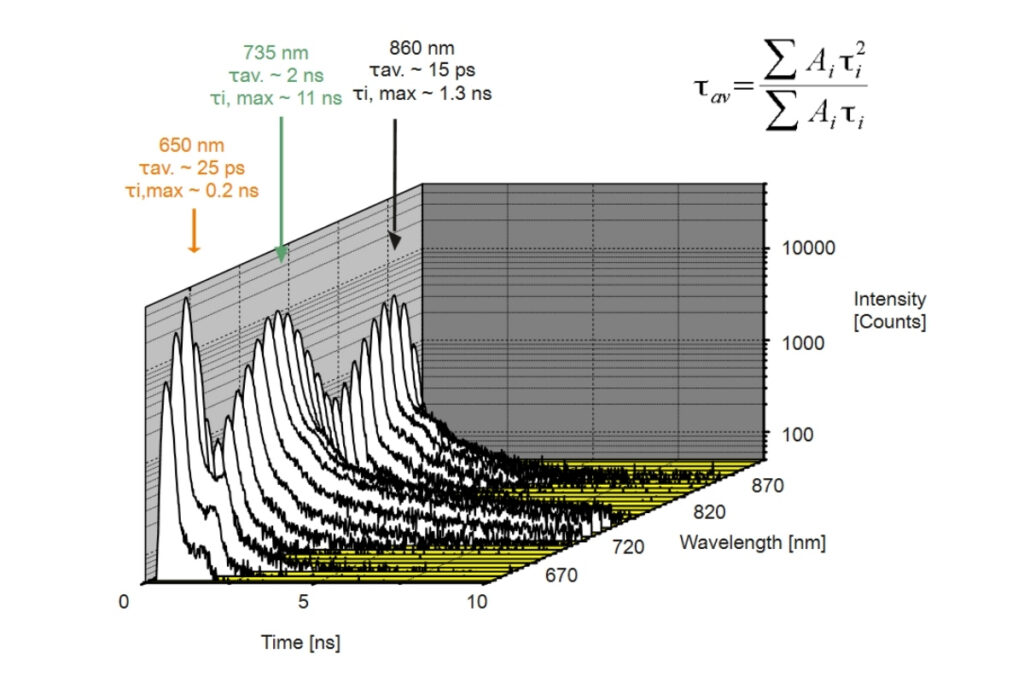
In TRPL imaging, the sample is excited by short laser pulses, producing photoluminescence that decays over time. The emitted photons are detected with picosecond temporal resolution and spatially assigned to image pixels. Time-Correlated Single Photon Counting (TCSPC) is commonly used to record photon arrival times relative to each excitation pulse. Repeating this process builds a decay histogram for every pixel, from which spatially resolved photoluminescence lifetime maps are reconstructed, revealing local variations in excited-state dynamics.
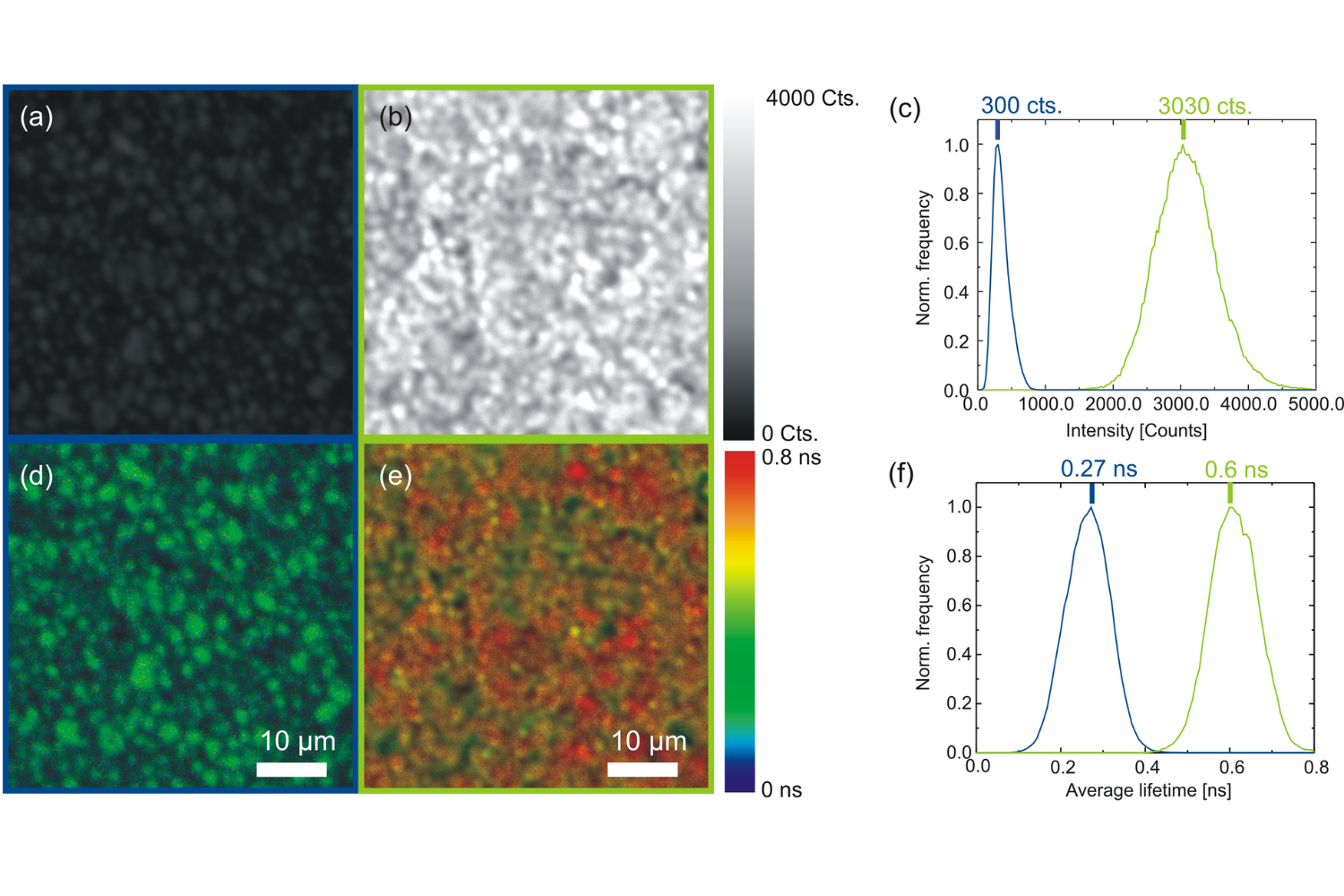
 SymphoTime 64: fluorescence lifetime imaging and correlation software.
TRPL imaging generates time-resolved photoluminescence decay curves for each image pixel. These decays are analyzed using monoexponential or multiexponential fitting to extract photoluminescence lifetimes and amplitude fractions. Lifetime maps visualize spatial variations in recombination dynamics, revealing defects, interfaces, or compositional heterogeneity. Fit-free methods such as intensity-weighted mean lifetimes or pattern-based classification facilitate rapid data interpretation. Quantitative TRPL imaging analysis enables direct comparison of local optoelectronic properties within complex materials.
PicoQuant’s SymphoTime 64 software enables intuitive TRPL Imaging data acquisition and decay analysis with integrated fitting in a single streamlined workflow.
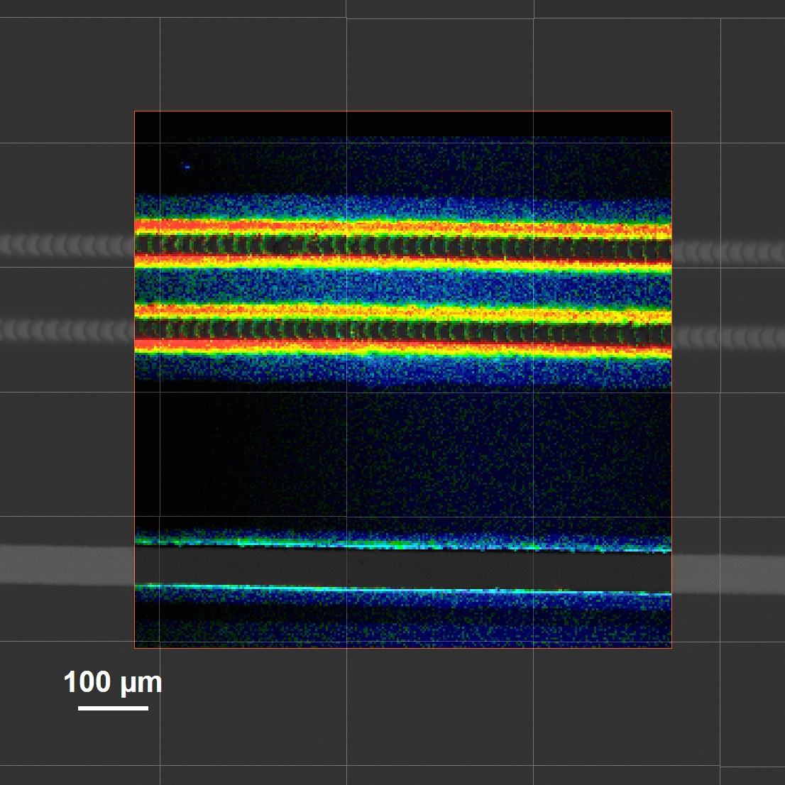
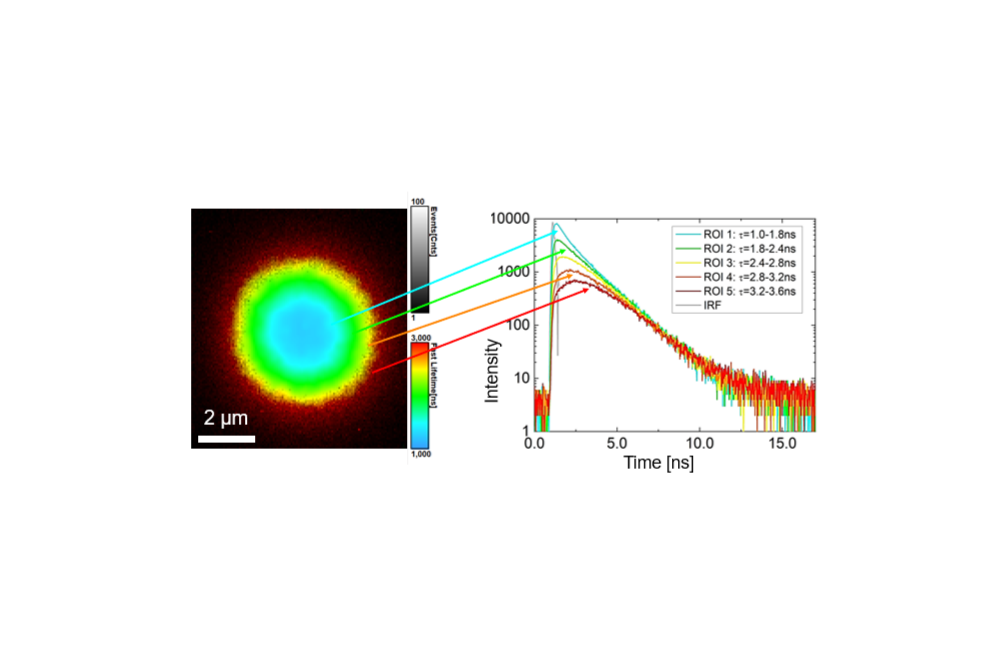 Carrier diffusion maps derived from time-resolved photoluminescence imaging. Decay curves from different regions of interest reveal spatial variations in recombination dynamics and enable extraction of diffusion-related parameters such as carrier diffusion length and diffusion coefficient.
TRPL imaging provides unique insight into spatially heterogeneous excited-state dynamics that cannot be accessed with bulk spectroscopy. By directly mapping photoluminescence lifetimes, the technique enables quantitative analysis of recombination pathways, defect-related losses, and charge transport processes. TRPL imaging is particularly valuable for correlating structural features with functional properties in advanced materials. It supports materials optimization by linking microscopic lifetime variations to composition, processing conditions, and device performance.
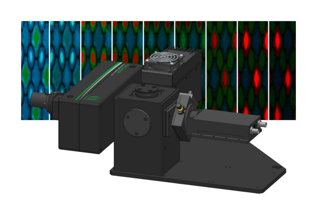
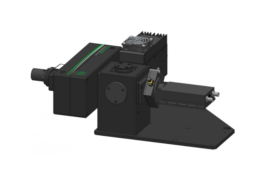
 Micro-PL upgrade combining a scanning microscope with a spectrometer for spatially resolved, time-resolved photoluminescence analysis.
Accurate TRPL imaging requires picosecond pulsed laser excitation, time-resolved single-photon detection, and precise synchronization electronics. High temporal resolution is essential to resolve fast photoluminescence decays, while stable scanning or imaging optics ensure spatial fidelity. Multi-channel TCSPC electronics enable efficient photon timing across image pixels. Equally important is the reliable optical and electronic communication between the spectrometer, microscope, and detection unit, as complex coupling and signal routing can strongly affect data quality. Together, these components form an integrated system capable of quantitative, spatially resolved photoluminescence lifetime imaging in materials science.
The following examples demonstrate how time-resolved photoluminescence imaging enables spatially resolved analysis of carrier dynamics, recombination processes, and material quality in advanced semiconductor systems.
Using the Solira time-resolved photoluminescence microscope, spatially resolved TRPL imaging reveals that laser-patterned regions in perovskite solar mini modules remain photoluminescent after laser structuring. Rather than completely removing the emissive material, the laser process modifies local photophysical properties and charge carrier dynamics. By combining localized TRPL measurements with spatial imaging in a single workflow, Solira enables a more comprehensive analysis of how device structuring affects semiconductor performance.
Time-resolved photoluminescence imaging enables non-destructive investigation of photovoltaic devices with high spatial resolution. By combining confocal microscopy with spectroscopic detection, carrier diffusion, power-dependent recombination dynamics, and lifetime heterogeneity can be analyzed across semiconductor structures. The approach supports quantitative correlation of structural inhomogeneities with photophysical behavior in CIGS and other thin-film solar cell materials.
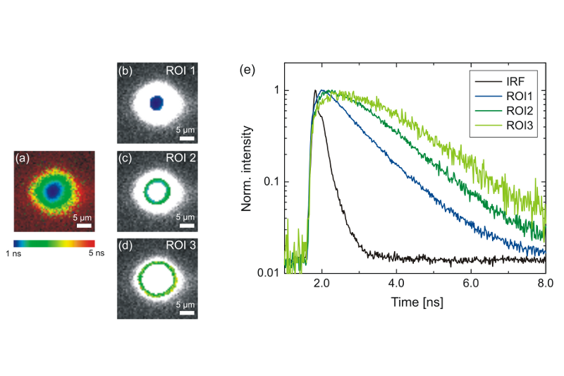
Time-resolved photoluminescence imaging of a GaAsP quantum well reveals spatially dependent carrier diffusion following localized excitation at 440 nm. Lifetime maps show increasing average lifetimes with radial distance from the excitation center, while decay analysis indicates diffusion-limited rise dynamics. Measurements were performed using an Olympus FluoView FV1000 equipped with PicoQuant’s LSM Upgrade Kit.
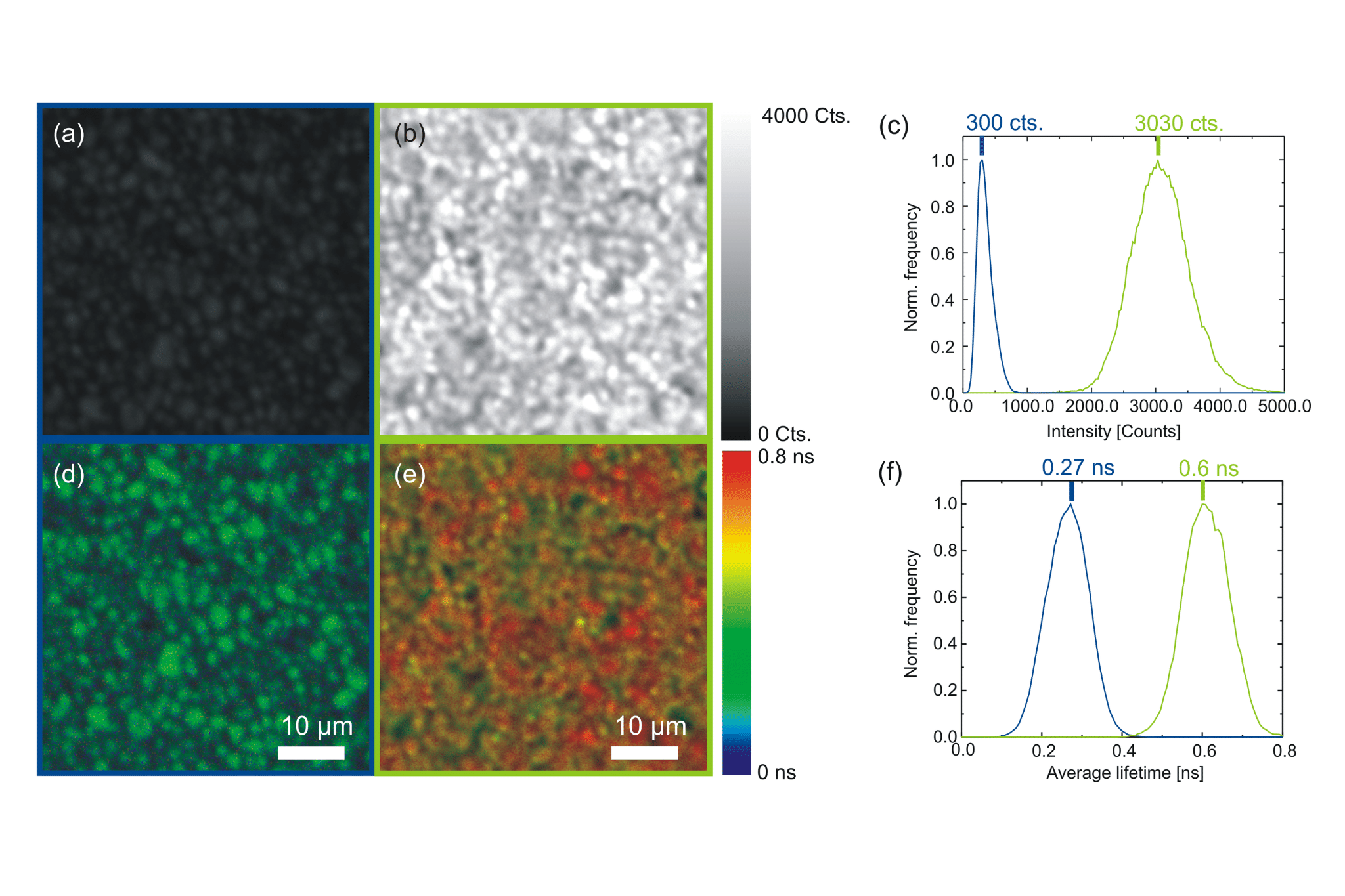
TRPL imaging of CdTe polycrystalline wafers before and after chloride-based thermal activation demonstrates significant increases in photoluminescence intensity and carrier lifetime. Lifetime maps and statistical distributions reveal enhanced recombination dynamics and spatial heterogeneity with millisecond acquisition times. Measurements were conducted using PicoQuant’s MicroTime 100 Time-Resolved Photoluminescence Microsope.
A steady-state optical imaging technique that maps the emission intensity of a material under continuous excitation. PL imaging provides spatial information on electronic states, defect-related transitions, and overall optical quality, but it lacks temporal resolution and cannot resolve excited-state dynamics.
A time-resolved spectroscopy technique that measures the temporal decay of photoluminescence following pulsed excitation. TRPL reveals recombination dynamics and excited-state lifetimes, but it lacks the spatial resolution achievable with TRPL imaging.