
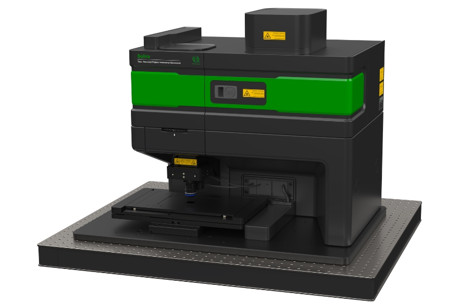
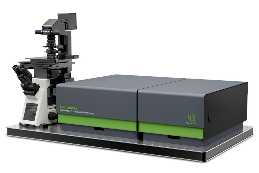
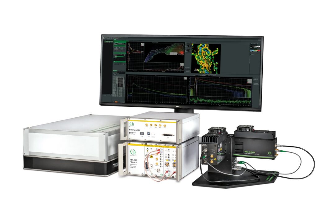
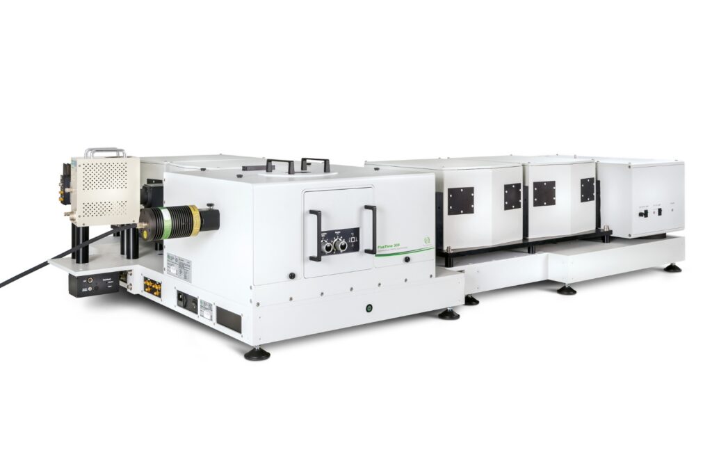
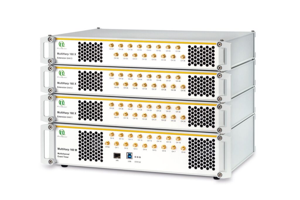
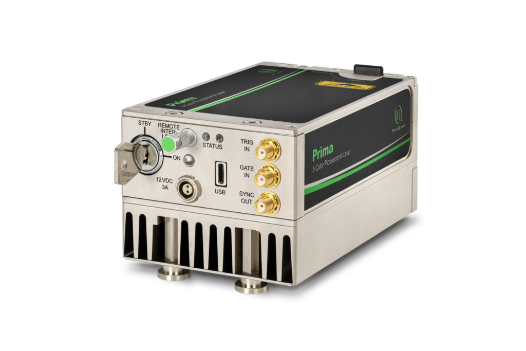

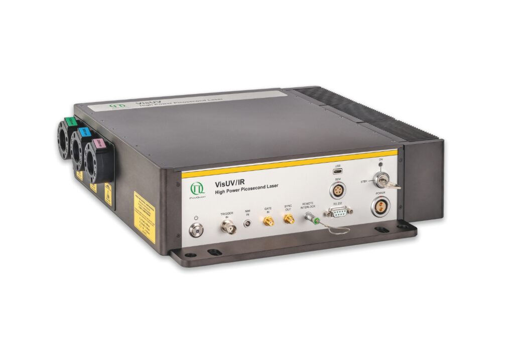

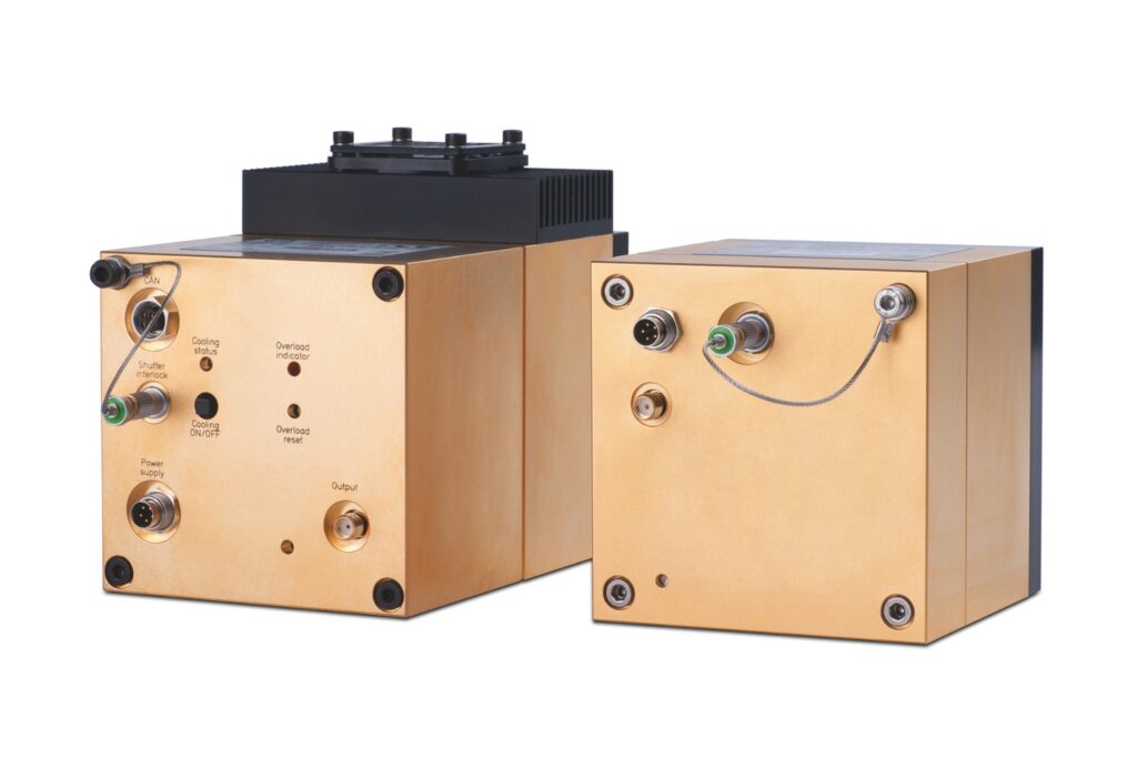
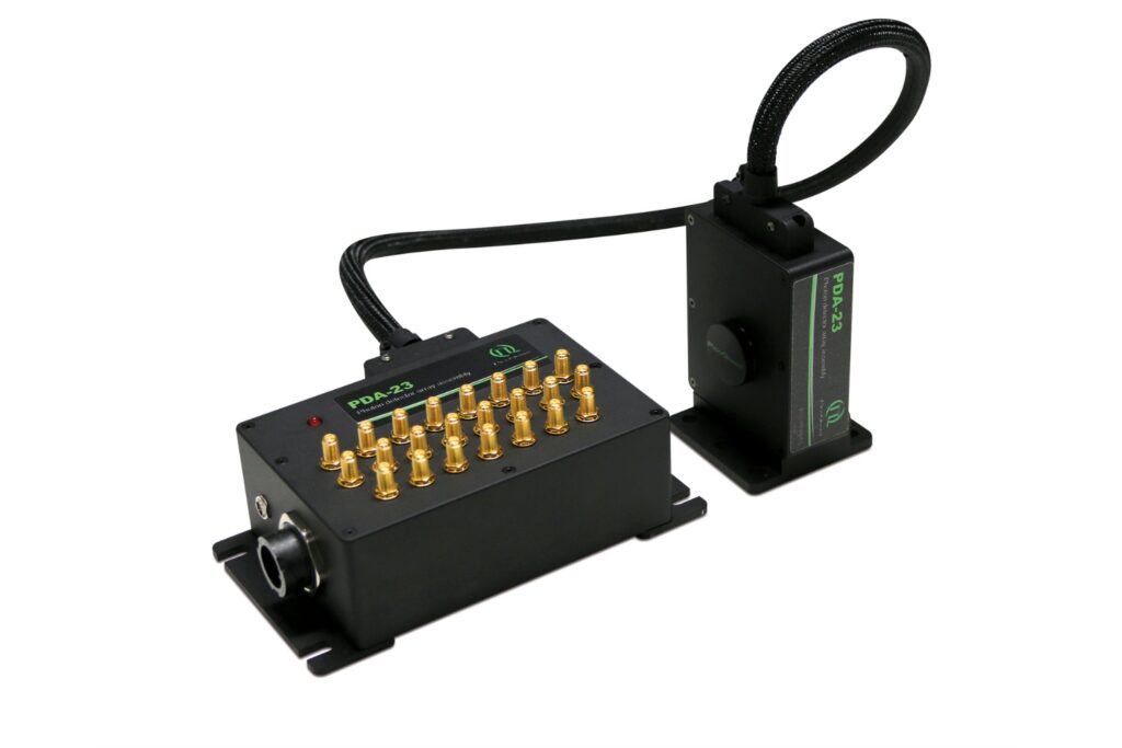
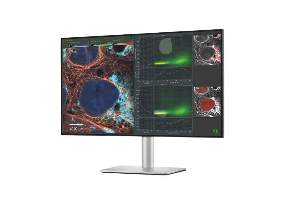
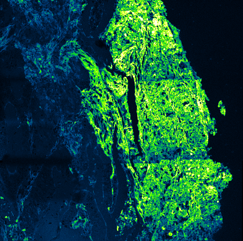
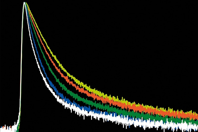


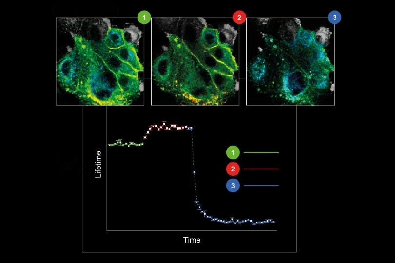
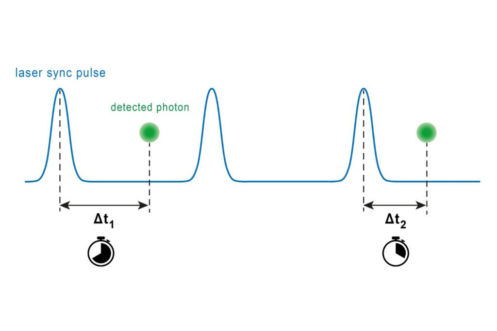
n a recently published paper, researchers at the Institute of Neurobiology at Heinrich Heine University Düsseldorf (Germany) have reported on a novel imaging method they developed in collaboration with colleagues from the University of Bonn (Germany) and PicoQuant. The method combines PicoQuant’s rapidFLIM approach – which allows recording fluorescence lifetime images at unprecedented speeds and temporal resolution – with multi-photon microscopy, offering high spatial resolution in the intact, highly scattering brain tissue.
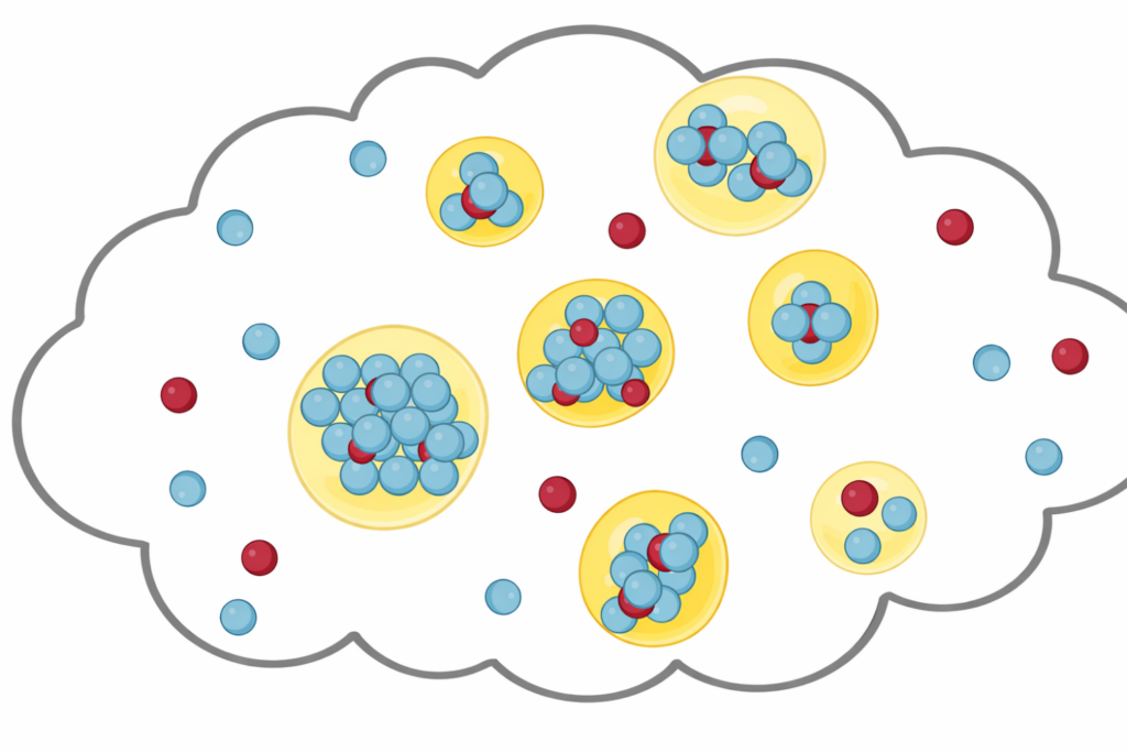
Thanks to this combination, the researchers were able to depict in real time the changes that occur when nerve cells are deprived of energy. They discovered that a breakdown in cellular energy supply – one of the most critical events during a stroke – leads to a rapid increase in sodium concentration inside of the cell and to subsequent swelling. A major discovery of this study is that a previously unknown mechanism involving TRPV4 ion channels plays a key role in the fatal sodium influx.
Link to publication: https://www.jneurosci.org/content/42/4/552/tab-article-info