header image news

News and Press Releases

April 18, 2024

New development enables rapid 3D FLIM imaging in living organoids

Combining single objective light-sheet microscopy with pulsed excitation and time-resolved SPAD array detection

Using a self-developed single objective light-sheet microscopy set-up, Valentin Dunsing-Eichenauer from Aix-Marseille Université & CNRS, Johan Hummert from PicoQuant, Ivan Michel Antolović from Pi Imaging Technology, and colleagues show lifetime-based multiplexing in 3D as well as time-lapse FLIM of mechanosensitive tension probes at record speed in living embryonic organoids. The set-up is based on pulsed excitation using a high power laser prototype and time-resolved detection on a newly available SPAD array detector.

Confocal FLIM is a popular tool for multiplexing and functional imaging of sensor fluorophores, but it is limited with regard to acquisition speed of large samples and live cell compatibility. Light-sheet microscopy, on the other hand, allows for fast and gentle imaging of large specimen. Combining both techniques opens new possibilities for fast volumetric FLIM.

Read the paper at bioRxiv >

New development enables rapid 3D FLIM imaging in living organoids

April 4, 2024

Enhanced synchronization capabilities with White Rabbit interface

Available for our multichannel event timers MultiHarp 150 and MultiHarp 160

The White Rabbit interface offers precise synchronization and timestamping for two separate time taggers with multiple channels. This ensures accurate time correlation between multiple devices or even different experiments, without cumbersome postprocessing. Amongst other applications, this is particularly relevant for quantum cryptography experiments.

PicoQuant's new Python wrapper snAPI allows you to easily implement White Rabbit into your experiments, enabling the simultaneous measurement initiation of two devices with predefinable measurement times. This ensures precise and reliable data acquisition, further streamlining synchronization capabilities across various applications.

White Rabbit interfaces are available with our MultiHarp 150 and MultiHarp 160 multichannel event timers.

Picture: Example of a network topology. The green bubbles stand for a MultiHarp 150 or MultiHarp 160 device. The blue bubbles represent the White Rabbit switches. 

Enhanced synchronization capabilities with White Rabbit interface

February 2, 2024

New application note:  Liquid-Liquid Phase Separation

Investigating the behavior of synapsin-1 in biological condensates with FLIM and FCS

Liquid-liquid phase separation (LLPS) is a physical phenomenon where a liquid containing several components demixes into two separate phases - a dense, condensed and a dilute phase - depending on the concentration and other stimuli. One key method to study LLPS is fluorescence microscopy, which allows researchers to visualize the formation and dynamics of condensates, and to observe the behavior of molecules inside condensates in real-time.

In our new application note our application specialist Maria Loidolt-Krüger describes how FLIM and multi-point FCS shed light on the behavior of synapsin-1 in biological condensates.

Download

New application note:  Liquid-Liquid Phase Separation

February 1, 2024

Course on Time-resolved Microscopy in Berlin

Registration is now open

From May 14 to 17, 2024 PicoQuant will hold the 16th International Course on "Time-resolved Microscopy and Correlation Spectroscopy" in Berlin. Benefit from this in-person event with theoretical as well as practical sessions, discussions, and networking opportunities. 

Register by February 19, 2024 and pay the early bird rate.

Course on Time-resolved Microscopy in Berlin

January 4, 2024

A deep dive into biomolecular condensates using single-molecule FRET

Webinar with Professor Samrat Mukhopadhyay

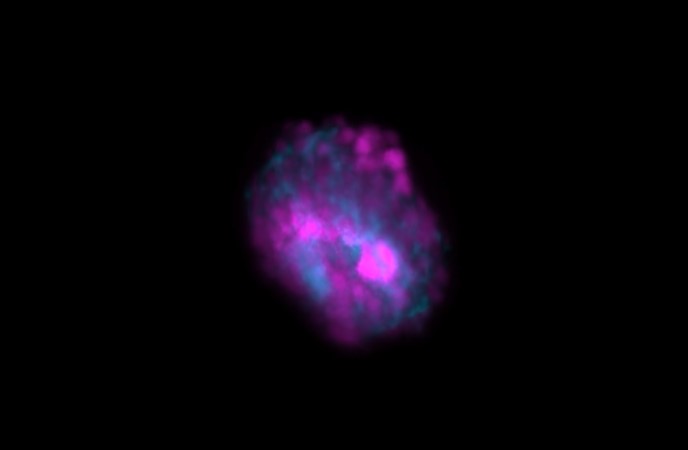
Cells house organelles enclosed by membranes, which partition cellular components. Recent captivating research suggests an additional means of intracellular compartmentalization and organization, namely liquid-liquid phase separation of proteins and nucleic acids, forming noncanonical membrane-less organelles. These dynamic biomolecular condensates, resembling liquids, may undergo undesirable irreversible phase transitions, leading to gel-like or solid-like amyloid aggregates. Such transformations are linked to various severe human diseases.

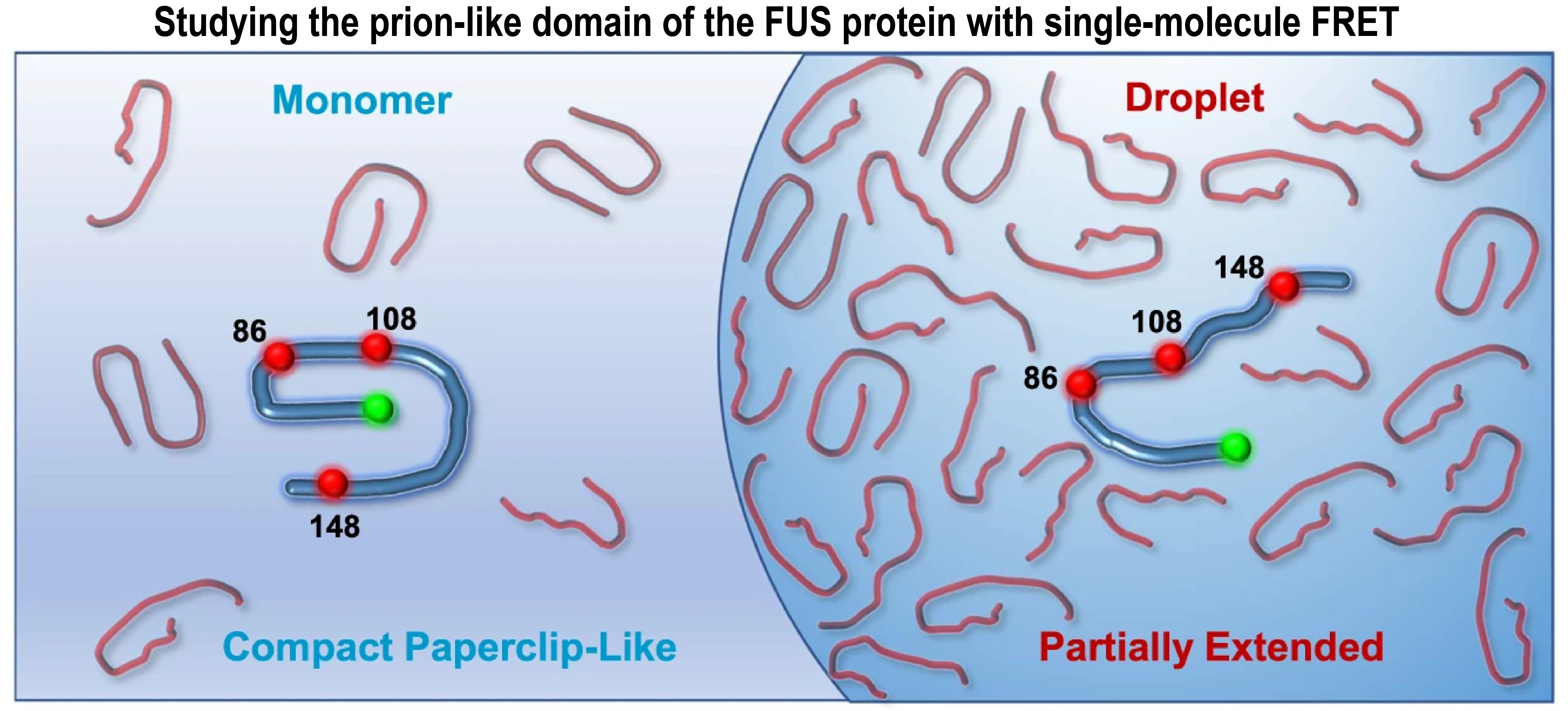
In a brand-new paper, Ashish Joshi and colleagues from the team of Samrat Mukhopadhyay at IISER Mohali take a close look at the molecular events during phase separation of the prion-like domain of the FUS protein with single-molecule fluorescence methods.

Our speaker, Professor Samrat Mukhopadhyay, will present ‘A deep dive into biomolecular condensates using single-molecule FRET’. The webinar is on Thursday 11 January at 14:00 GMT, 15:00 CET and 19:30 IST.

If you want to learn what single molecule fluorescence microscopy techniques revealed about the complex behavior of the FUS low-complexity domain during liquid-liquid phase separation, register for our upcoming webinar featuring Prof. Samrat Mukhopadhyay, co-hosted with FocalPlane.

Register!

A deep dive into biomolecular condensates using single-molecule FRET